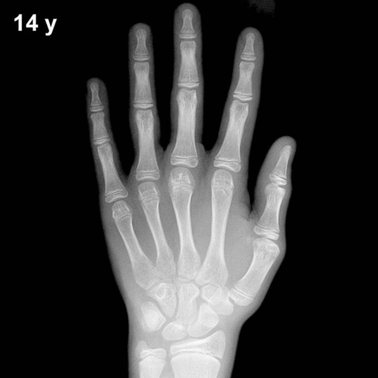
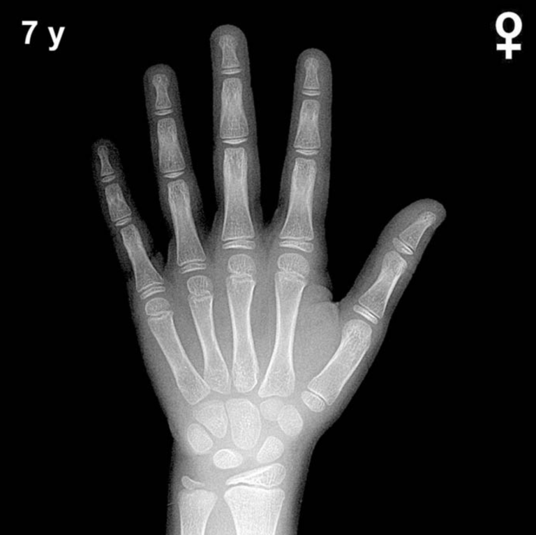
Bone Age in Boys Aged 5.5 Years — Greulich-Pyle Hand and Wrist X-Ray Reference
Bone age assessment using a left-hand and wrist radiograph is a cornerstone of pediatric endocrine and growth disorder evaluation. The Greulich-Pyle (GP) method compares a child’s skeletal maturation against standardized atlas plates derived from a North American reference population. In boys aged 5.5 years, this assessment helps identify deviations consistent with growth hormone deficiency, precocious puberty, hypothyroidism, or constitutional growth delay.
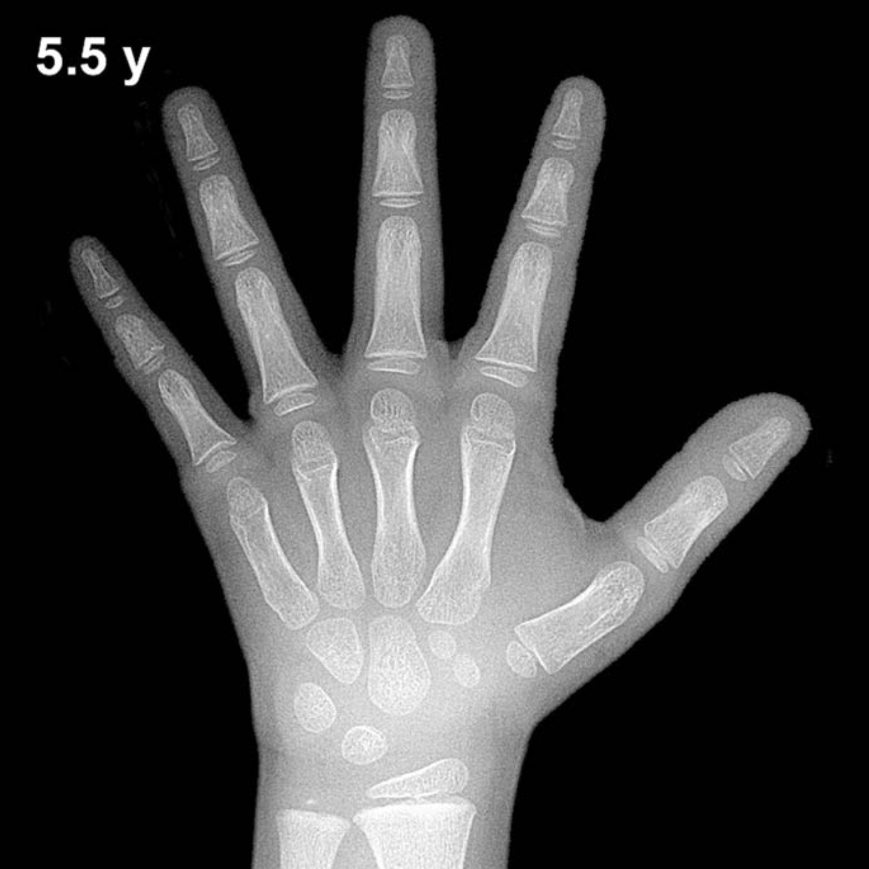
Expected Ossification Centers and Skeletal Findings
By 5.5 years of age in boys, most primary carpal ossification centers are well established. The capitate and hamate are among the earliest to appear (typically by 3–6 months of life) and should be well-ossified and clearly defined by this age. The triquetral ossification center, which typically emerges between 2–3 years, is expected to be present and progressively enlarging. The lunate, appearing around 3–4 years in boys, should also be visible and growing in size.
At 5.5 years, the scaphoid, trapezium, and trapezoid ossification centers are typically appearing or recently established, generally within the 4–6 year window in boys. The distal radial epiphysis, present from approximately 1 year of age, should be well-formed and broadening. The distal ulnar epiphysis typically appears between 5–7 years, so its presence or absence at this exact age is a key landmark to note. Metacarpal and proximal phalangeal epiphyses are present and maturing, with progressive increase in their relative width compared to shaft diameter.
- Capitate & Hamate: Well-ossified (present since infancy)
- Triquetral: Present and enlarging
- Lunate: Present, progressive ossification
- Scaphoid, Trapezium, Trapezoid: Typically present or just appearing
- Distal radial epiphysis: Well-formed and broadening
- Distal ulnar epiphysis: May be just appearing (~5–7 yr window)
- Pisiform: Not yet expected (typically 11–14 yr in boys)
Clinical Pearls
Bone age in boys at 5.5 years carries a standard deviation of approximately ±1 year, meaning skeletal ages between roughly 4.5 and 6.5 years may fall within the normal range. Girls are skeletally ahead of boys by approximately 6–12 months at this age. A bone age advanced by more than 2 SD should prompt evaluation for precocious puberty, congenital adrenal hyperplasia, or exogenous androgen exposure. Conversely, a delayed bone age of more than 2 SD raises concern for growth hormone deficiency, hypothyroidism, or constitutional delay of growth and puberty. A key pitfall is over-reliance on a single carpal center’s appearance; skeletal maturation should be assessed globally across multiple bones to avoid misclassification.
Reference: Greulich WW, Pyle SI. Radiographic Atlas of Skeletal Development of the Hand and Wrist. 2nd ed. Stanford University Press, 1959.