Bone Age in Boys Aged 17 Years — Greulich-Pyle Hand and Wrist X-Ray Reference
Bone age assessment using a left-hand and wrist radiograph is a cornerstone of pediatric endocrine and growth evaluation, providing an estimate of skeletal maturity independent of chronological age. The Greulich-Pyle method compares the patient's radiograph against standardized atlas plates to assign a skeletal age. At 17 years in boys, the assessment is particularly important for evaluating late-pubertal growth, constitutional delay, and estimating remaining growth potential.
Expected Ossification Centers and Skeletal Findings
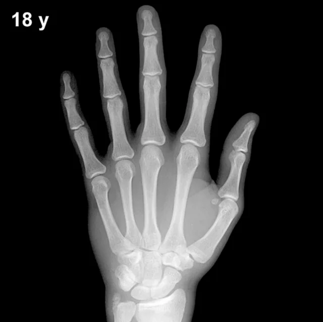
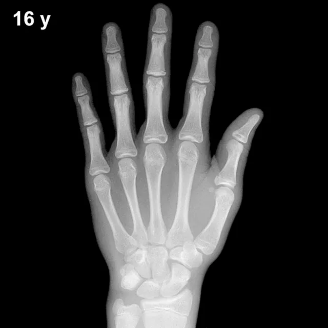
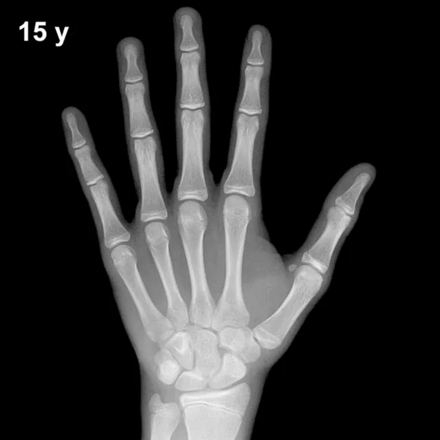
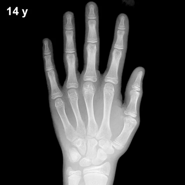
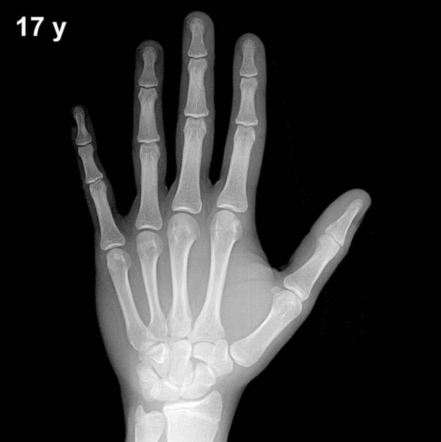
By 17 years of age in boys, virtually all ossification centers of the hand and wrist are present and well developed. All eight carpal bones — capitate, hamate, triquetral, lunate, scaphoid, trapezium, trapezoid, and pisiform — are typically ossified and approach mature morphology. The pisiform, which appears relatively late (around 11–14 years in boys), is well established by this age.
A defining skeletal feature at this stage is the progressive fusion of epiphyses to their respective metaphyses. The distal radial and ulnar epiphyses, present since early childhood, are typically in an advanced stage of fusion or may show complete fusion in skeletally mature individuals. Epiphyseal fusion of the phalanges generally proceeds from distal to proximal; the distal phalangeal epiphyses often fuse first, followed by middle and proximal phalangeal epiphyses. The metacarpal epiphyses are also typically fusing or fused by this age.
- Carpal bones: All eight present; mature or near-mature morphology expected.
- Distal radius and ulna: Epiphyses in advanced fusion or completely fused.
- Phalangeal epiphyses: Fusion progressing or complete, particularly at distal and middle phalanges.
- Sesamoid of the thumb: Fully ossified; typically appears peripubertally (around 13–15 years in boys) and is well established by 17 years.
- Metacarpal epiphyses: Fusing or fused in skeletally advanced individuals.
Clinical Pearls
At 17 years, the standard deviation for bone age in boys spans approximately ±10–12 months, meaning a skeletal age of roughly 15.5–18+ years may still fall within a broad normal range. Girls' skeletal maturation typically runs approximately 1–2 years ahead of boys at equivalent chronological ages, a difference that narrows as full maturity approaches. A bone age significantly advanced beyond 17 years in a boy suggests early epiphyseal fusion and reduced residual growth potential, as seen in conditions such as precocious puberty or exogenous androgen exposure. Conversely, a notably delayed bone age may point toward constitutional delay of growth and puberty, hypogonadism, growth hormone deficiency, or hypothyroidism. A key interpretive pitfall at this age is over-relying on a single radiograph without correlating pubertal staging (Tanner stage) and growth velocity data, as epiphyseal fusion timing varies considerably between individuals. Reference: Greulich WW, Pyle SI. Radiographic Atlas of Skeletal Development of the Hand and Wrist. 2nd ed. Stanford University Press, 1959.