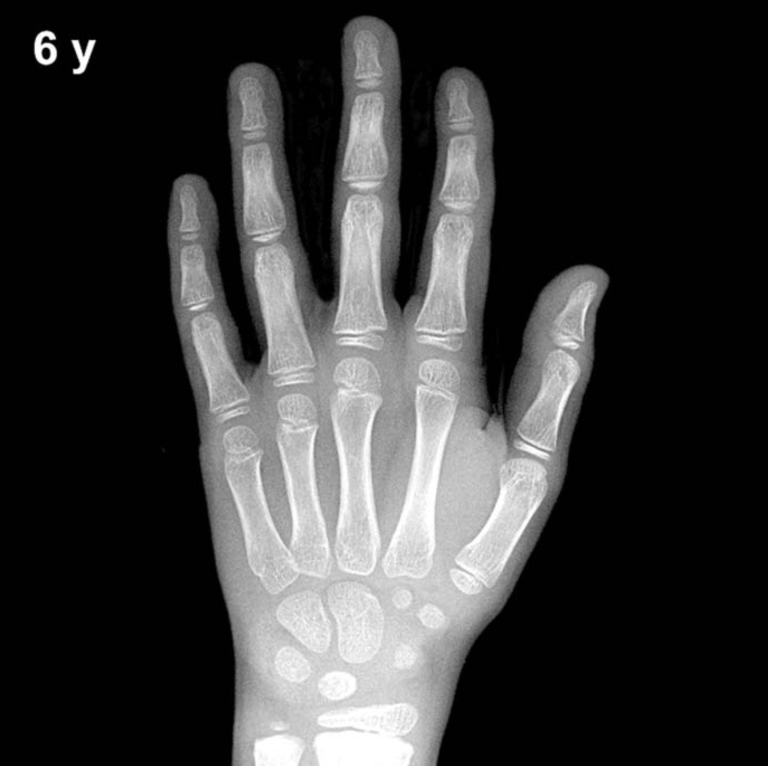
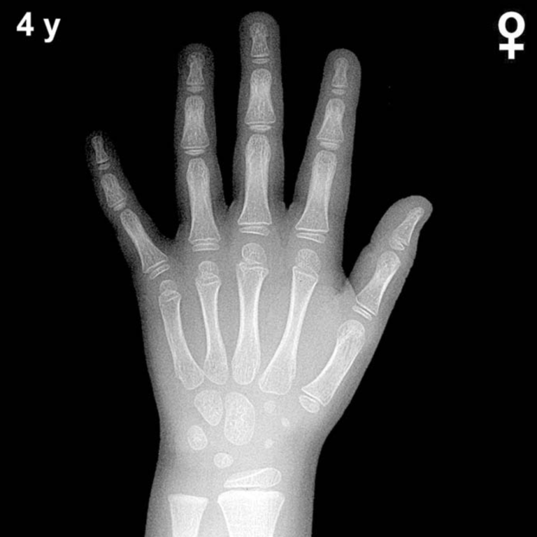
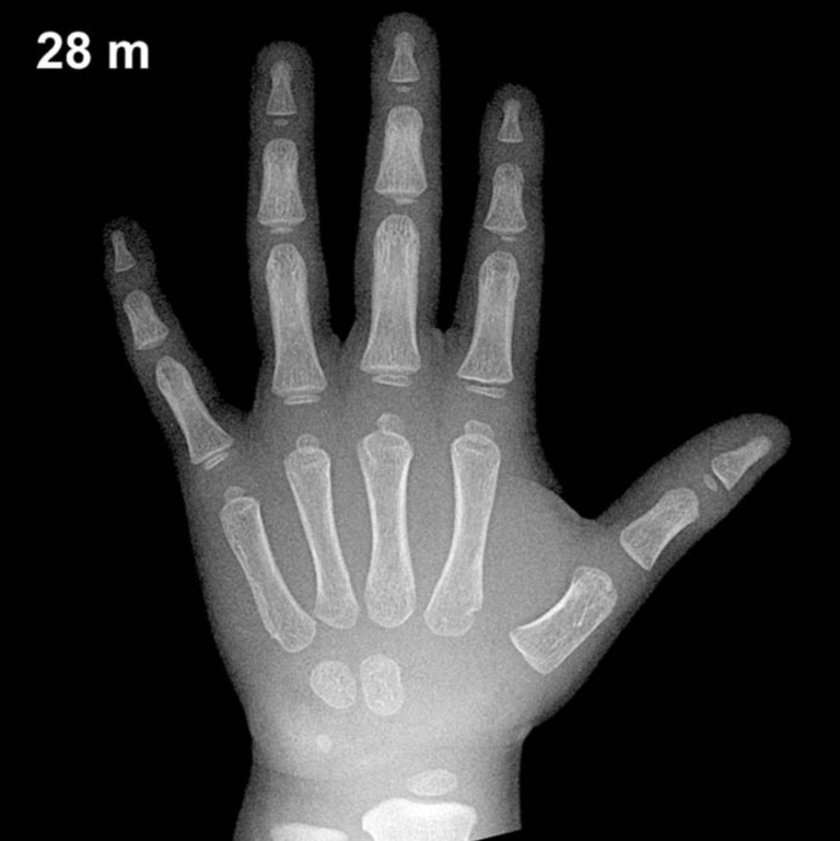
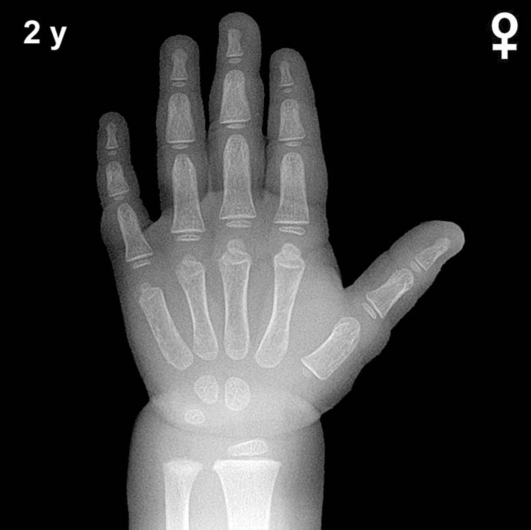
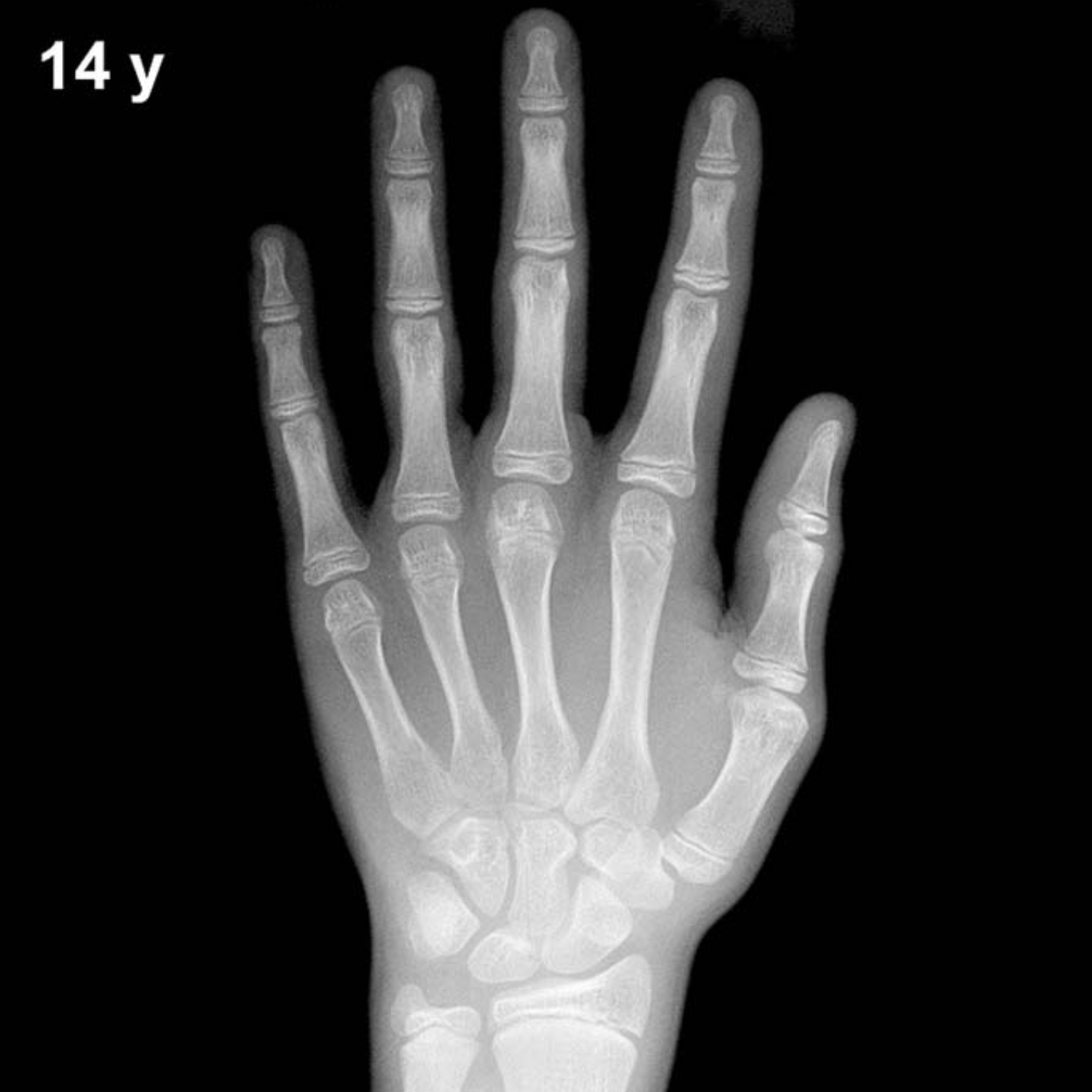
Bone Age in Boys Aged 14 Years — Greulich-Pyle Hand and Wrist X-Ray Reference
Bone age assessment using a left hand and wrist radiograph is a cornerstone of pediatric endocrine and growth evaluation. The Greulich-Pyle (GP) method compares a patient’s skeletal maturation against standard atlas plates derived from healthy North American children. In 14-year-old boys, this assessment is particularly valuable for evaluating pubertal timing, growth hormone adequacy, and conditions such as constitutional delay of growth and puberty.
Expected Ossification Centers and Skeletal Findings
By 14 years in boys, all major carpal ossification centers are well established. The capitate and hamate (present from infancy), triquetral, lunate, scaphoid, trapezium, trapezoid, and pisiform should all be visible and demonstrating progressive maturation. The pisiform, which typically appears between approximately 11–14 years in boys, should be present or emerging by this age and serves as a useful pubertal landmark.
Epiphyseal maturation is the dominant feature at this age. The distal radial and ulnar epiphyses are well developed, with the distal radial epiphysis showing increasing width and density. Epiphyses of the metacarpals and proximal, middle, and distal phalanges are broadening and beginning to cap their respective metaphyses. The adductor sesamoid of the thumb, a reliable marker of mid-puberty in boys, typically appears around Greulich-Pyle bone age 13–14 years and should be present or just appearing at this chronological age.
- All 8 carpal bones: ossified and maturing in morphology
- Pisiform: present by this age in most boys
- Thumb sesamoid: typically visible at GP bone age ~13–14 years in boys
- Distal radius/ulna epiphyses: well formed, progressive capping of metaphyses
- Phalangeal and metacarpal epiphyses: broadening; partial to complete capping expected
Clinical Pearls
A normal standard deviation of approximately ±1–1.5 years is accepted in GP bone age assessment at this age. Girls’ skeletal maturation characteristically leads boys’ by roughly 1–2 years, so the GP standard plates for boys and girls must never be interchanged. A bone age significantly advanced beyond 14 years in a boy should prompt evaluation for gonadotropin-dependent or gonadotropin-independent precocious puberty, congenital adrenal hyperplasia, or exogenous androgen exposure. Conversely, a notably delayed bone age raises concern for growth hormone deficiency, hypothyroidism, hypogonadism, or constitutional delay of growth and puberty — the latter being by far the most common cause and typically a diagnosis of exclusion.
A key interpretive pitfall is population variability: the GP atlas was standardized on a predominantly white, mid-20th-century American cohort, and skeletal maturation norms may differ across ethnicities. Bone age should always be interpreted alongside clinical history, pubertal staging, growth velocity, and auxological data rather than as an isolated finding. Reference: Greulich WW, Pyle SI. Radiographic Atlas of Skeletal Development of the Hand and Wrist. 2nd ed. Stanford University Press, 1959.