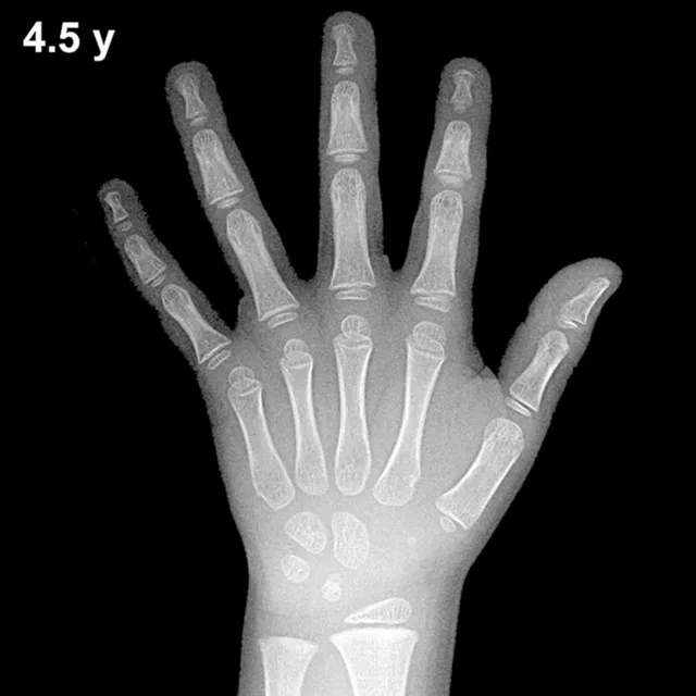
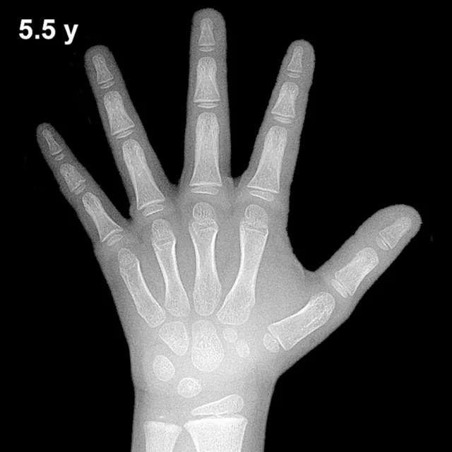
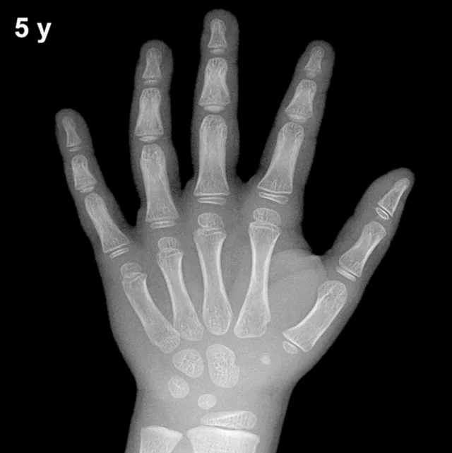
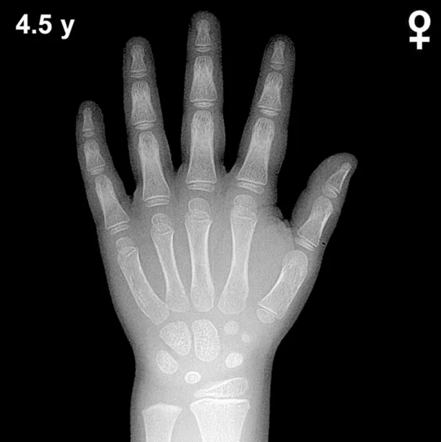
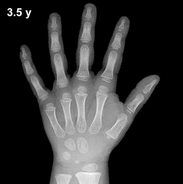
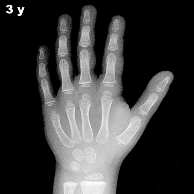
Bone Age in Boys Aged 4 Years — Greulich-Pyle Hand and Wrist X-Ray Reference
Bone age assessment using a left hand and wrist radiograph is a standard method for evaluating skeletal maturity in children. The Greulich-Pyle method compares a child's radiograph against standardized atlas plates to determine whether skeletal development is appropriate for chronological age. In 4-year-old boys, this assessment is particularly relevant in the workup of growth disorders, precocious or delayed puberty, and endocrine conditions.
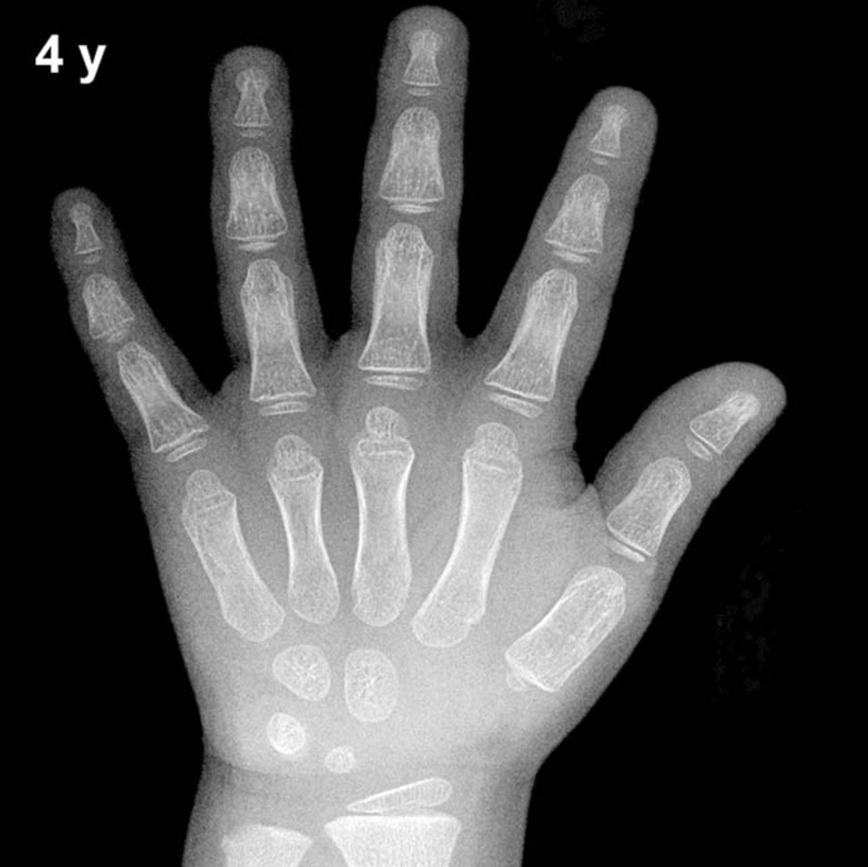
Expected Ossification Centers and Skeletal Findings
By 4 years of age in boys, several carpal and epiphyseal ossification centers are typically well established. The capitate and hamate are among the earliest carpal bones to ossify (approximately 3 and 6 months of age, respectively) and should be clearly visible. The triquetral ossification center, which typically appears between 2 and 3 years, is generally present by this age. The lunate, expected around 3–4 years, may be newly visible or in the early stages of ossification in boys at this age.
The distal radial epiphysis is well ossified by 4 years, having typically appeared around 1 year of age. Epiphyses of the metacarpals and proximal, middle, and distal phalanges should all be present and show progressive development. The scaphoid, trapezium, and trapezoid are not reliably expected until approximately 4–6 years, so their absence or early appearance at this age is within normal variation. The distal ulnar epiphysis typically appears between 5 and 7 years and is generally not yet ossified at age 4 in boys.
- Capitate: present (ossifies ~3 months)
- Hamate: present (ossifies ~6 months)
- Triquetral: typically present by age 4
- Lunate: may be early or newly visible (~3–4 years)
- Distal radial epiphysis: well established
- Scaphoid/trapezium/trapezoid: absent to early (expected ~4–6 years)
- Pisiform and distal ulnar epiphysis: not yet expected
Clinical Pearls
Skeletal maturation in girls typically runs 6–12 months ahead of boys at this age, so atlas plates for 4-year-old boys reflect a slightly less advanced pattern than those for girls of the same chronological age. A bone age more than 2 standard deviations above the mean may suggest precocious puberty, exogenous androgen exposure, or congenital adrenal hyperplasia. Conversely, a significantly delayed bone age at this age warrants consideration of growth hormone deficiency, hypothyroidism, or constitutional delay of growth and puberty.
A key pitfall is over-reliance on a single ossification center for age assignment; the Greulich-Pyle method requires assessment of the overall skeletal pattern across all visible bones. Ethnic and nutritional variation can also influence skeletal maturity independent of endocrine pathology. Reference: Greulich WW, Pyle SI. Radiographic Atlas of Skeletal Development of the Hand and Wrist. 2nd ed. Stanford University Press, 1959.