Bone Age in Boys Aged 6 Years — Greulich-Pyle Hand and Wrist X-Ray Reference
Bone age assessment using a left hand and wrist radiograph is a cornerstone of pediatric endocrine and growth evaluation. The Greulich-Pyle (GP) method compares a child’s skeletal maturation against sex-specific standard radiographs, providing an estimated skeletal age independent of chronological age. In 6-year-old boys, this assessment is commonly requested in the workup of growth concerns, precocious or delayed puberty, and constitutional growth delay.
Expected Ossification Centers and Skeletal Findings
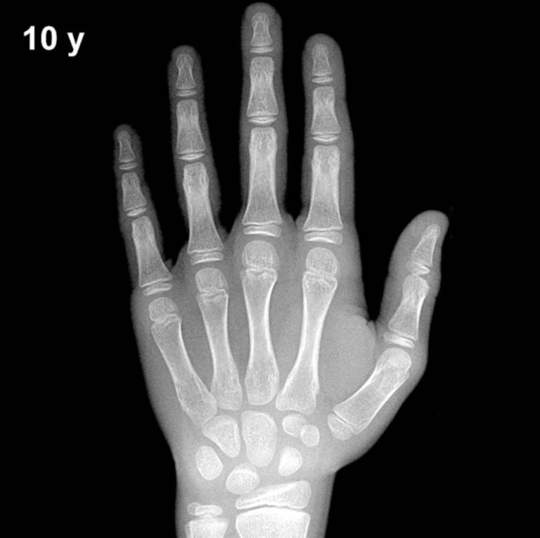
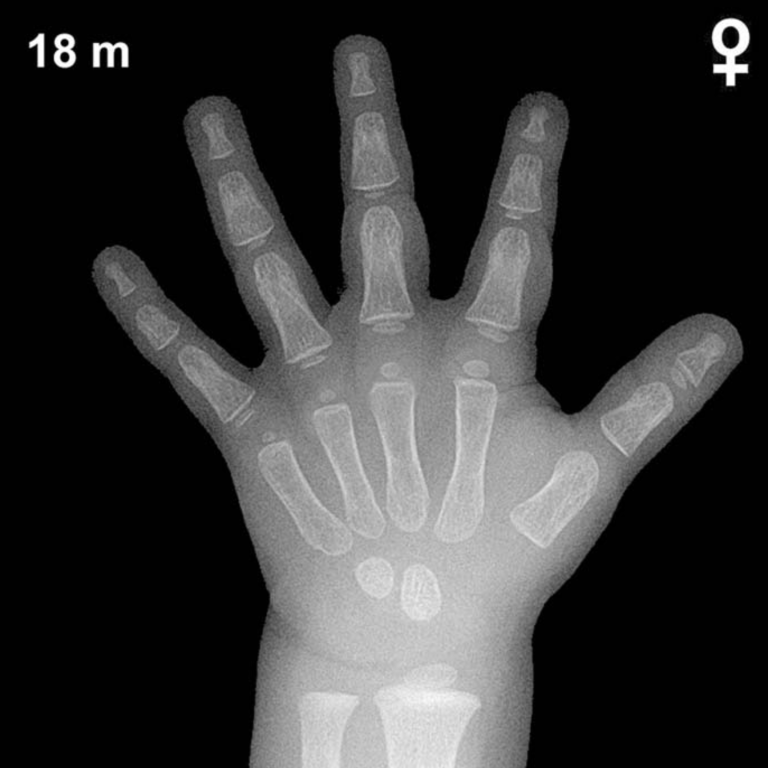
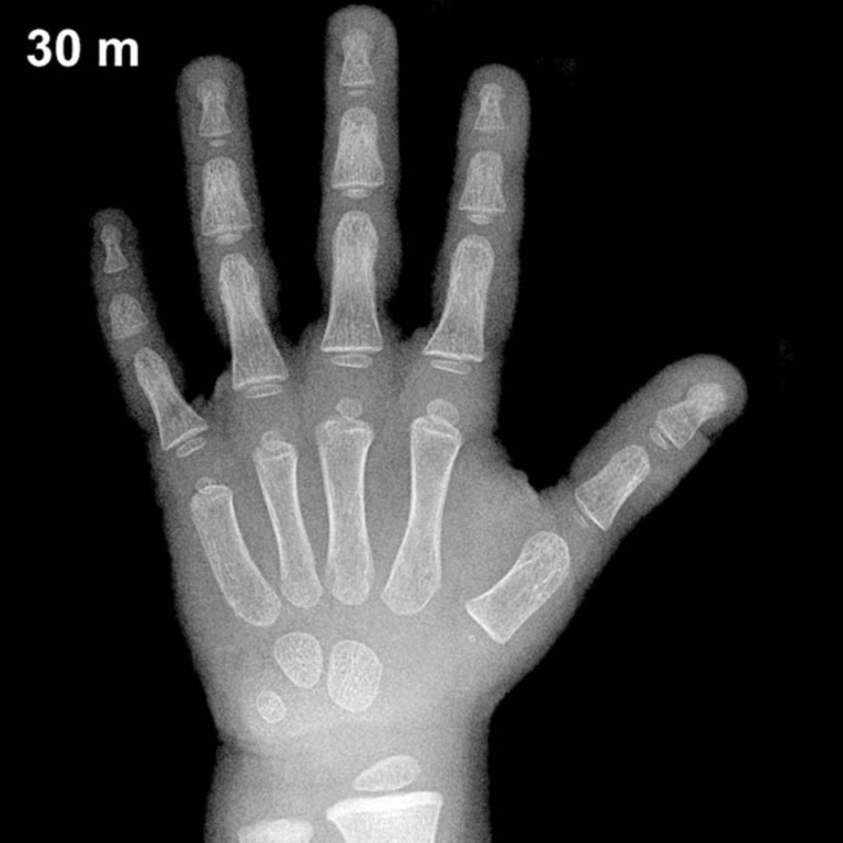
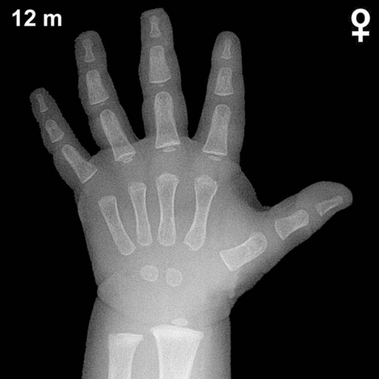
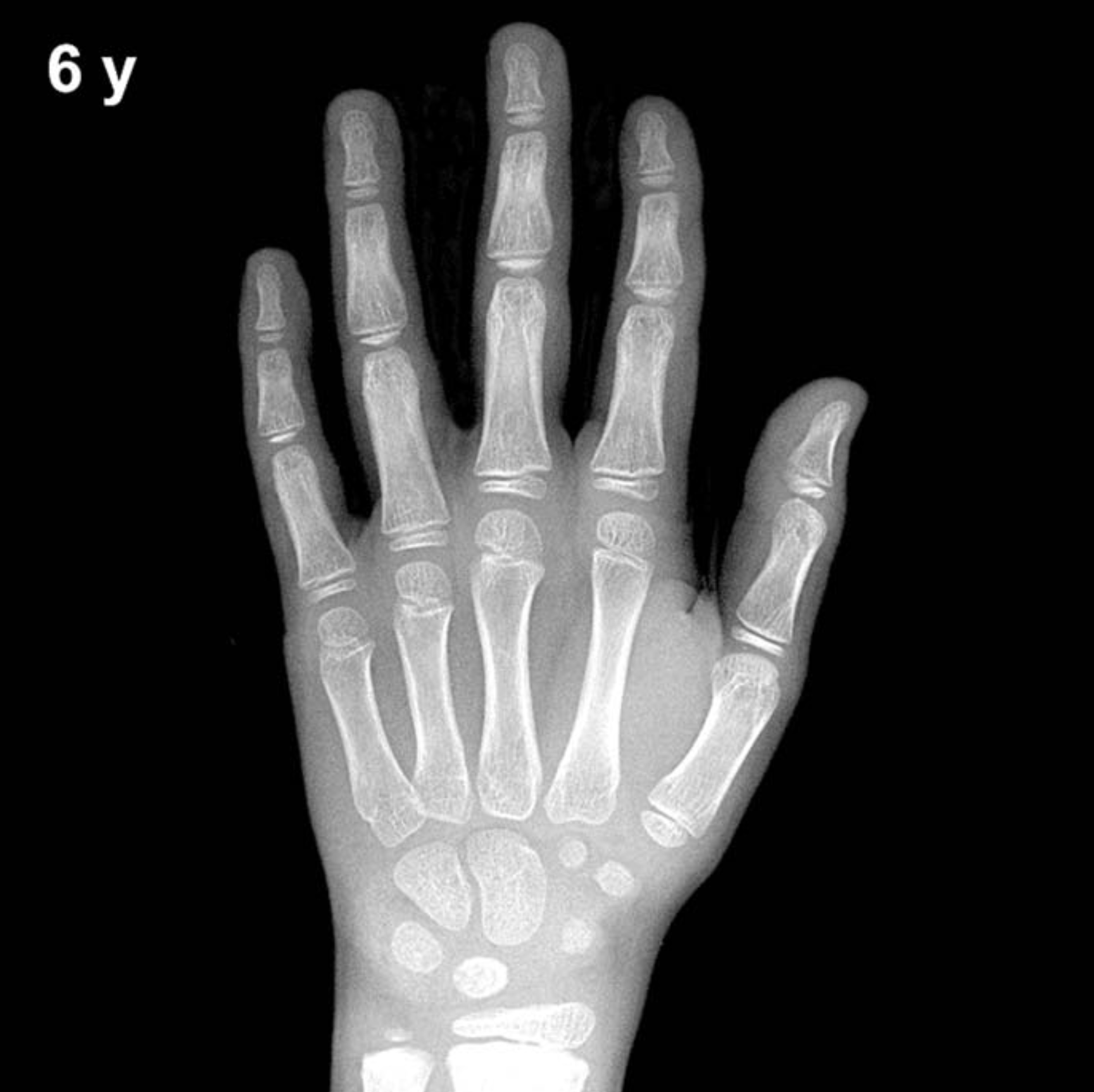
By 6 years of age in boys, all primary carpal ossification centers are typically visible. The capitate and hamate (appearing around 3 and 6 months of age, respectively) are well established and should demonstrate mature, well-corticated morphology. The triquetral (typically appearing around 2–3 years) and lunate (around 3–4 years) are expected to be clearly present. The scaphoid, trapezium, and trapezoid — which typically appear between 4 and 6 years — may be present or just emerging at this age in boys, and their appearance is an important skeletal milestone to evaluate carefully.
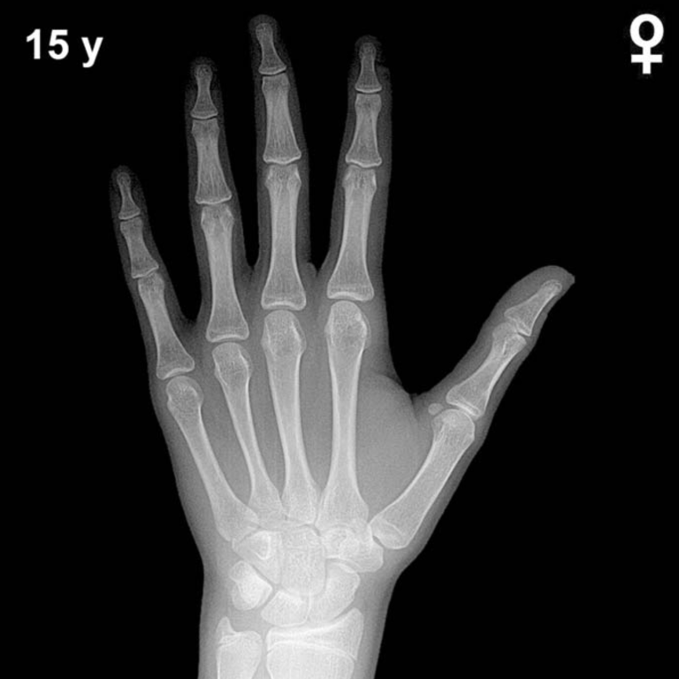
The distal radial epiphysis, typically ossifying around 1 year of age, should be well developed by age 6. The distal ulnar epiphysis, which generally appears between 5 and 7 years, may be visible but is often just appearing in boys at this age. Epiphyses of the metacarpals and phalanges should be present and show progressive widening relative to their diaphyses. The pisiform is not yet expected in boys at age 6, typically appearing between 11 and 14 years. The thumb sesamoid is also not yet anticipated, as it is a peripubertal landmark.
Clinical Pearls
Skeletal maturation in girls is consistently ahead of boys by approximately 1–2 years during middle childhood, meaning the GP standard plates for a 6-year-old boy reflect a somewhat less mature skeleton than an age-matched girl. The standard deviation for bone age at this chronological age is approximately ±1 year, so a bone age of 5–7 years would fall within the expected range. A bone age advanced by more than 2 years should prompt evaluation for precocious puberty, adrenal hyperplasia, or exogenous androgen exposure. A bone age delayed by more than 2 years raises concern for growth hormone deficiency, hypothyroidism, or constitutional delay of growth and puberty.
A key interpretive pitfall is over-reliance on a single ossification center; skeletal maturation should be assessed holistically across multiple bones. Racial and nutritional factors may also influence maturation and should be considered when the GP atlas — derived from a mid-20th-century North American reference population — is applied to diverse patient groups. Reference: Greulich WW, Pyle SI. Radiographic Atlas of Skeletal Development of the Hand and Wrist. 2nd ed. Stanford University Press, 1959.