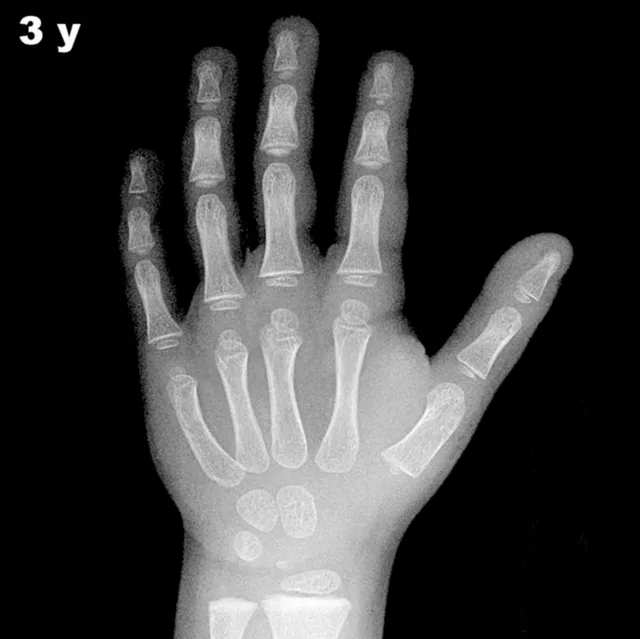
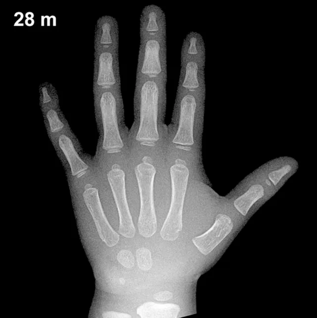
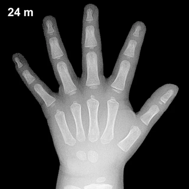
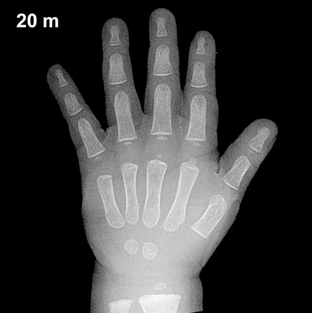
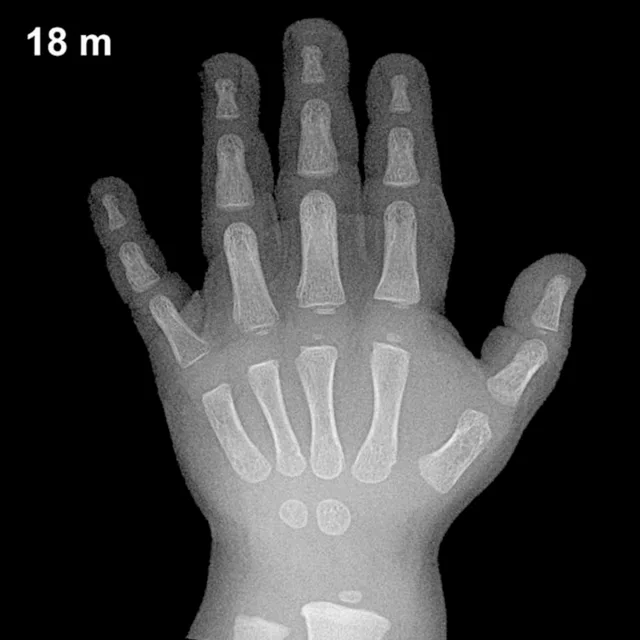
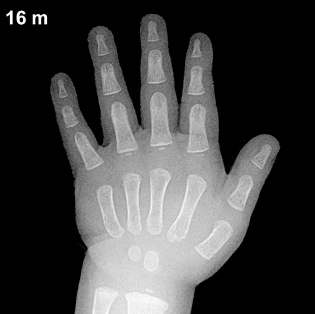
Bone Age in Boys Aged 30 Months — Greulich-Pyle Hand and Wrist X-Ray Reference
Bone age assessment uses a hand and wrist radiograph to estimate skeletal maturity, comparing ossification centers against standardized atlas references. The Greulich-Pyle method remains the most widely used technique in clinical practice for evaluating growth disorders, endocrine conditions, and, when necessary, forensic age estimation. In boys aged 30 months, accurate bone age interpretation provides a valuable baseline for identifying skeletal advancement or delay relative to chronological age.
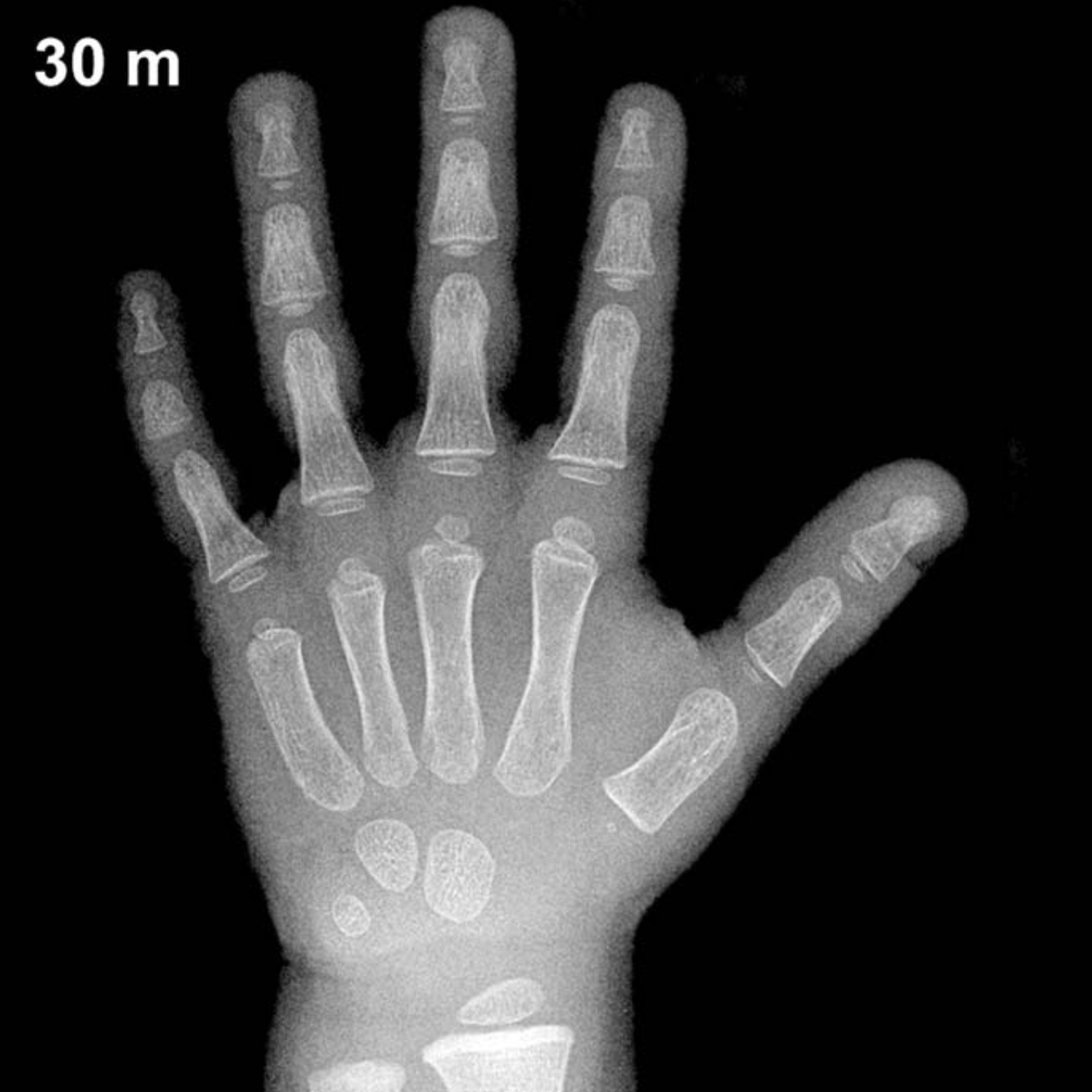
Expected Ossification Centers and Skeletal Findings
By 30 months of age in boys, several carpal and epiphyseal centers are typically visible on a hand and wrist radiograph. The capitate and hamate are among the earliest carpal bones to ossify, appearing around 3 and 6 months respectively, and should be well established by this age. The triquetral ossification center typically emerges between 2 and 3 years in boys and may be present or just appearing at 30 months. The lunate generally appears somewhat later, around 3 to 4 years, and is not consistently expected at this age.
The distal radial epiphysis is typically visible by approximately 1 year of age and should be clearly ossified at 30 months. The remaining carpal centers — scaphoid, trapezium, and trapezoid — are not expected until approximately 4 to 6 years of age in boys, and the pisiform ossifies considerably later, typically between 11 and 14 years in boys. The distal ulnar epiphysis generally appears between 5 and 7 years and would not be expected at this age. Epiphyseal development of the metacarpals and phalanges continues to progress gradually through early childhood.
Clinical Pearls
Skeletal maturation in girls is known to run ahead of boys by approximately 1 to 2 years throughout childhood, a difference already measurable in early toddlerhood. At 30 months, normal bone age in boys may show variability of roughly ±6 months around the chronological age. A bone age significantly advanced for a 30-month-old boy may raise concern for precocious puberty or exposure to excess androgens, while a notably delayed bone age may suggest growth hormone deficiency, hypothyroidism, or constitutional delay of growth and puberty. A key interpretive pitfall at this age is overestimating carpal ossification variability; the number and size of ossified centers can differ substantially between healthy children, and isolated findings should always be correlated with clinical and endocrine data before drawing diagnostic conclusions.
Reference: Greulich WW, Pyle SI. Radiographic Atlas of Skeletal Development of the Hand and Wrist. 2nd ed. Stanford University Press, 1959.