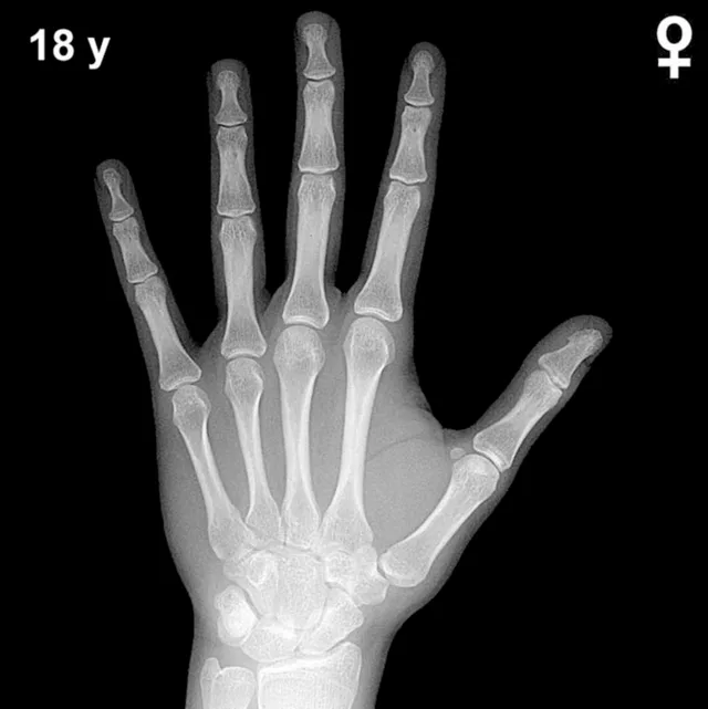
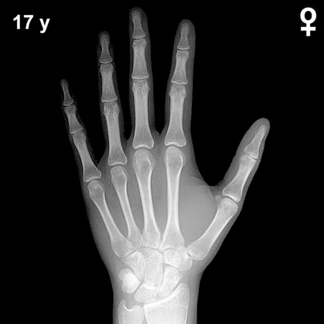
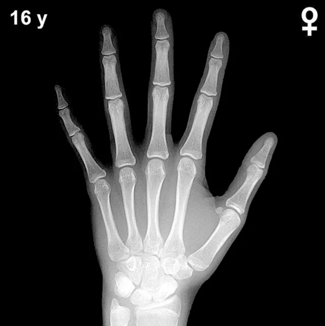
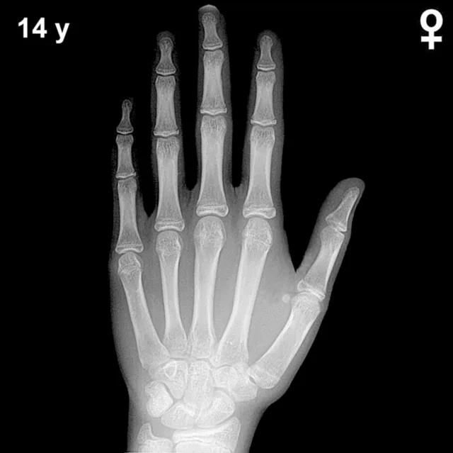
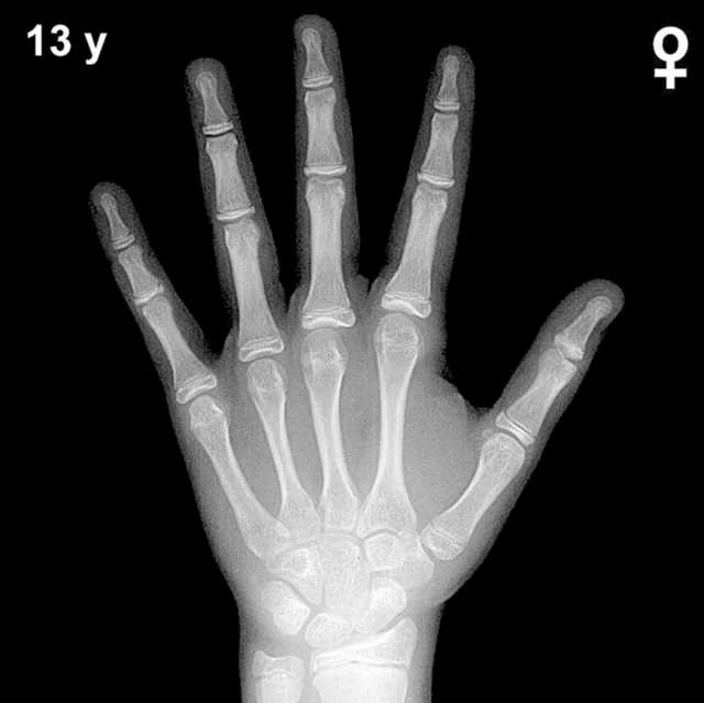
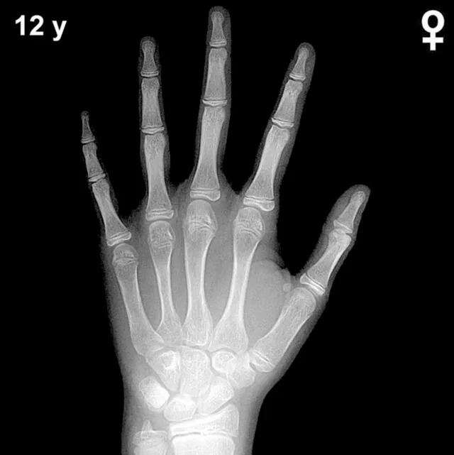
Bone Age in Girls Aged 15 Years — Greulich-Pyle Hand and Wrist X-Ray Reference
Bone age assessment using the Greulich-Pyle atlas compares a child's left-hand and wrist radiograph against standardized reference plates to estimate skeletal maturity. In girls aged 15 years, this evaluation is particularly relevant for investigating precocious or delayed puberty, monitoring growth disorders, and supporting endocrine workup. Accurate skeletal age determination helps clinicians estimate remaining growth potential and guides therapeutic decisions.
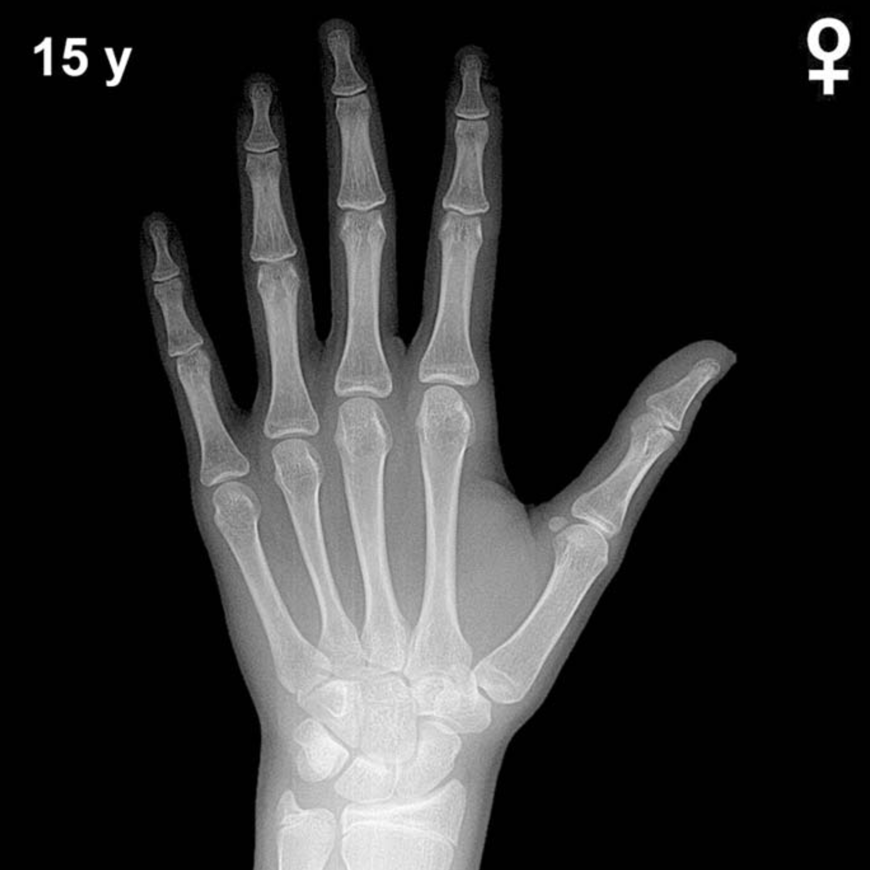
Expected Ossification Centers and Skeletal Findings
By 15 years in girls, skeletal maturation is advanced and approaching completion. All eight carpal bones — capitate, hamate, triquetral, lunate, scaphoid, trapezium, trapezoid, and pisiform — are well ossified and should be clearly visible. The pisiform, which typically appears between 9–12 years in girls, is well established by this age. Carpal bone morphology is essentially adult in configuration, with mature cortical margins and trabecular patterns.
The epiphyses of the distal radius and distal ulna are typically fusing or show significant fusion at this age in girls. Epiphyseal fusion at the distal radius generally begins around 14–16 years in girls and may be complete or near-complete by 15 years in more skeletally advanced individuals. The metacarpal and phalangeal epiphyses are similarly in advanced stages of fusion. The sesamoid of the thumb, a key peripubertal marker, is fully ossified and should be well developed. Epiphyseal-diaphyseal fusion across the hand is largely complete or nearing completion, indicating late adolescent skeletal maturity consistent with a Greulich-Pyle bone age of approximately 15 years.
Clinical Pearls
At 15 years, girls' skeletal maturity is typically ahead of boys of the same chronological age by approximately 1.5–2 years. A bone age significantly advanced beyond 15 years in a girl may suggest a history of precocious puberty or exposure to excess sex steroids, while a notably delayed bone age warrants evaluation for conditions such as growth hormone deficiency, hypothyroidism, Turner syndrome, or constitutional delay of growth and puberty. At this late stage of skeletal development, the standard deviation of bone age estimates narrows, but inter-reader variability remains a recognized pitfall — particularly when distinguishing near-complete from complete epiphyseal fusion, which can be subtle on standard radiographs.
A key interpretive pitfall is over-reliance on a single skeletal landmark; assessment should integrate findings across all visible epiphyses and carpal bones rather than focusing on any one site. Ethnic variation in skeletal maturation rates, not accounted for in the original Greulich-Pyle reference population, should also be considered when interpreting results in diverse populations. Reference: Greulich WW, Pyle SI. Radiographic Atlas of Skeletal Development of the Hand and Wrist. 2nd ed. Stanford University Press, 1959.