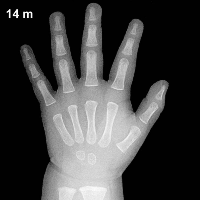
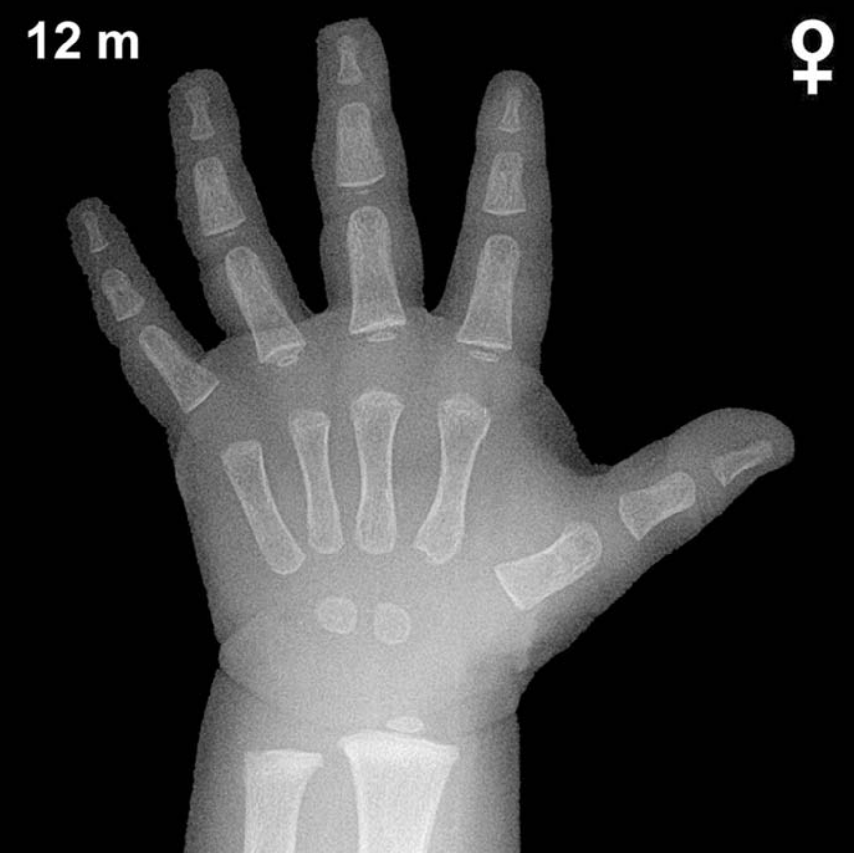
Bone Age in Girls Aged 12 Months — Greulich-Pyle Hand and Wrist X-Ray Reference
Bone age assessment using a left-hand and wrist radiograph is a cornerstone of pediatric skeletal maturity evaluation, most commonly performed using the Greulich-Pyle (GP) atlas. In infants and toddlers, it plays an important role in identifying early growth disturbances, evaluating endocrine disorders such as hypothyroidism or growth hormone deficiency, and providing forensic age estimation. At 12 months of age in girls, only a limited number of ossification centers are expected, making each visible center highly significant.
Expected Ossification Centers and Skeletal Findings
At 12 months, the skeletal maturity of girls is notably more advanced than that of age-matched boys. The capitate and hamate are typically the first carpal bones to ossify, appearing around 3 and 6 months respectively, and should be consistently present by 12 months in girls. The distal radial epiphysis typically appears around 9–12 months in girls and is expected to be visible or just emerging at this age, representing an important landmark at this stage.
The remaining carpal bones are not yet expected: the triquetral typically appears between 2–3 years, the lunate between 3–4 years, the scaphoid, trapezium, and trapezoid between 4–6 years, and the pisiform much later at approximately 9–12 years in girls. The distal ulnar epiphysis is generally not visible until approximately 5–7 years of age. Metacarpal and proximal phalangeal epiphyses may be just beginning to emerge at this age in girls.
- Capitate: Present (ossifies ~3 months)
- Hamate: Present (ossifies ~6 months)
- Distal radial epiphysis: Appearing or present (~9–12 months in girls)
- All other carpals: Not yet expected at 12 months
Clinical Pearls
There is meaningful inter-individual variability in skeletal maturity even among healthy infants; the GP atlas standards carry a standard deviation of approximately ±2–3 months at this age. Girls are skeletally ahead of boys by several months during infancy, a difference that widens through childhood. A bone age significantly advanced beyond 12 months in a girl should prompt consideration of precocious puberty or exogenous androgen/estrogen exposure, while a markedly delayed bone age raises concern for hypothyroidism, growth hormone deficiency, or nutritional deficiency. A key pitfall at this age is over-relying on a single ossification center: the distal radial epiphysis can be difficult to visualize due to positioning or exposure, and its apparent absence should be interpreted cautiously before concluding skeletal delay.
Reference: Greulich WW, Pyle SI. Radiographic Atlas of Skeletal Development of the Hand and Wrist. 2nd ed. Stanford University Press, 1959.