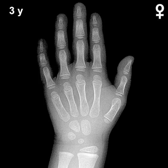
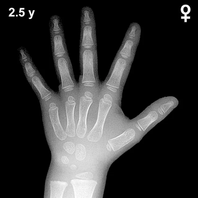
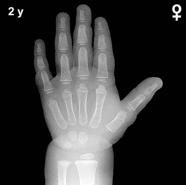
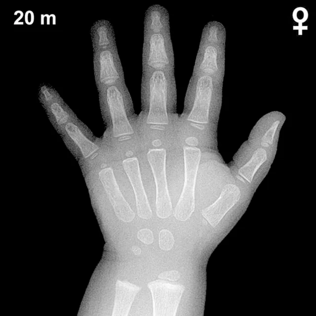
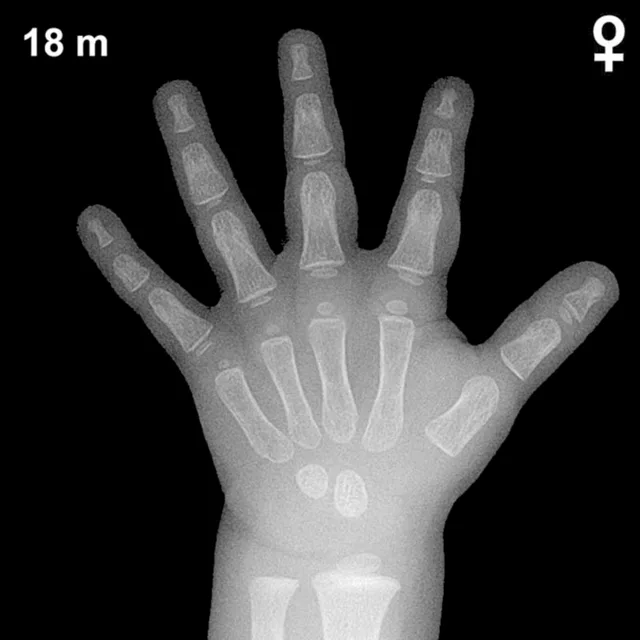
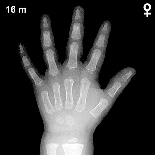
Bone Age in Girls Aged 28 Months — Greulich-Pyle Hand and Wrist X-Ray Reference
Bone age assessment using a left hand and wrist radiograph is a standard tool for evaluating skeletal maturation in children. The Greulich-Pyle method compares the radiograph to sex-specific atlas standards to determine whether skeletal development is concordant with chronological age. In girls aged 28 months, this assessment is particularly useful in the workup of growth faltering, suspected endocrine disorders, or precocious puberty.
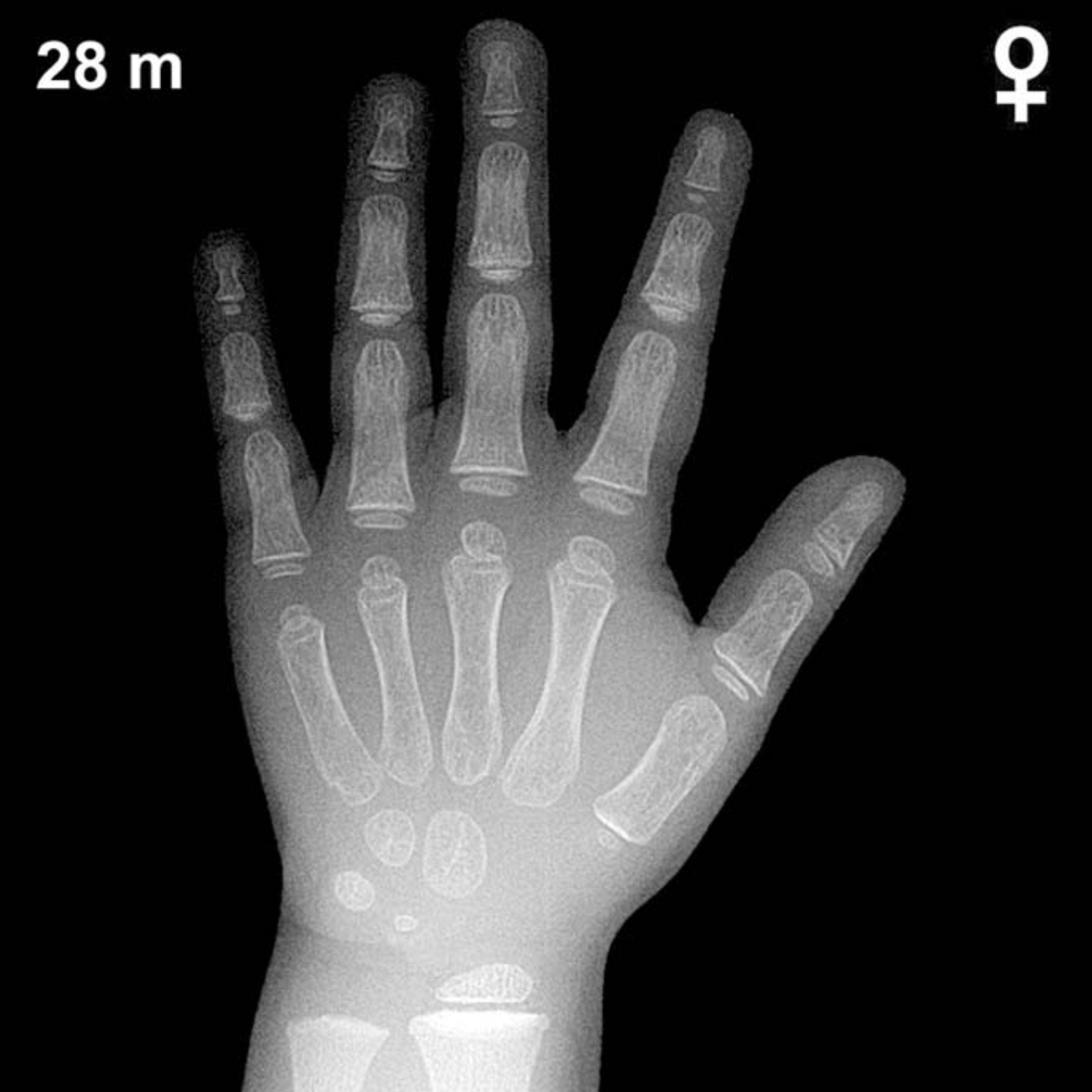
Expected Ossification Centers and Skeletal Findings
By approximately 28 months of age in girls, several carpal and epiphyseal ossification centers are typically present. The capitate and hamate are among the earliest carpals to ossify, appearing around 3 and 6 months respectively, and should be well established by this age. The triquetral ossification center typically emerges between 2 and 3 years in girls and may be visible or just appearing at 28 months. The lunate center generally appears between 3 and 4 years and is not yet expected at this age.
The distal radial epiphysis is typically present by the end of the first year and should appear as a well-defined ossification center by 28 months. The distal ulnar epiphysis is not yet expected, as it generally appears between 5 and 7 years. Epiphyses of the metacarpals and proximal, middle, and distal phalanges should be ossifying progressively, with phalangeal epiphyses becoming increasingly defined during the second and third years of life.
- Present by 28 months (girls): Capitate, hamate, distal radial epiphysis, metacarpal and phalangeal epiphyses (developing)
- Emerging around this age: Triquetral ossification center
- Not yet expected: Lunate, scaphoid, trapezium, trapezoid, pisiform, distal ulnar epiphysis
Clinical Pearls
Girls are typically skeletally advanced compared to boys of the same chronological age, often by several months during early childhood. At 28 months, a bone age that is significantly advanced (more than 2 standard deviations) may raise concern for precocious puberty or excess androgen or growth hormone exposure, whereas a notably delayed bone age may suggest hypothyroidism, growth hormone deficiency, or constitutional delay of growth and development. Clinicians should interpret bone age in the context of growth velocity, pubertal staging, and relevant laboratory findings rather than as an isolated finding.
A key pitfall at this age is the normal variability in carpal ossification timing; the triquetral, in particular, shows wide individual variation in its appearance. Attributing significance to its absence at 28 months without supporting clinical data is generally unwarranted. Reference: Greulich WW, Pyle SI. Radiographic Atlas of Skeletal Development of the Hand and Wrist. 2nd ed. Stanford University Press, 1959.