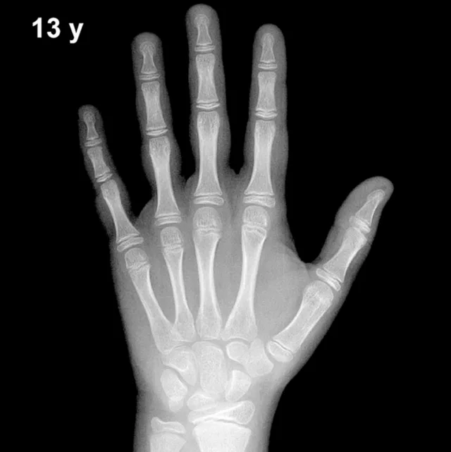
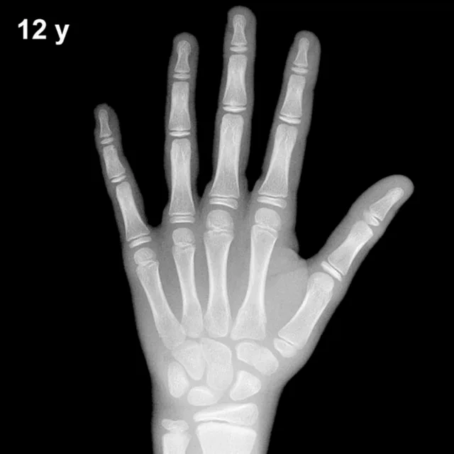
Bone Age in Boys Aged 18 Years — Greulich-Pyle Hand and Wrist X-Ray Reference
Bone age assessment using a left hand and wrist radiograph is a fundamental tool in pediatric radiology, allowing clinicians to estimate skeletal maturity independently of chronological age. The Greulich-Pyle method compares the patient's radiograph against standardized atlas plates derived from a North American reference population. At 18 years in boys, this assessment is particularly relevant for evaluating delayed puberty, growth hormone deficiency, and for medicolegal age estimation.
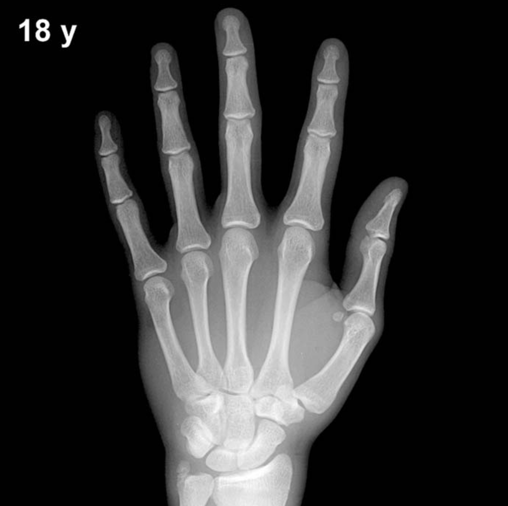
Expected Ossification Centers and Skeletal Findings
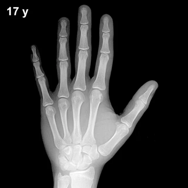
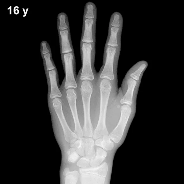
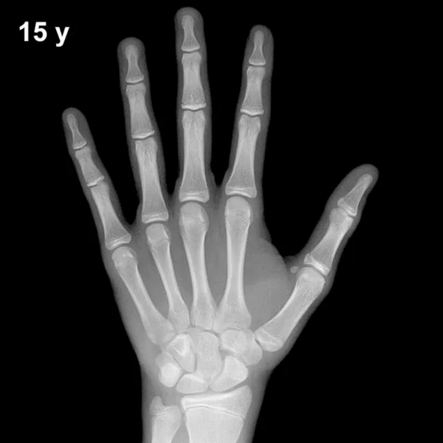
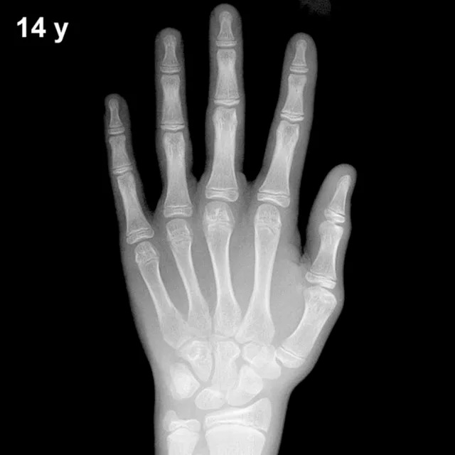
By 18 years of age in boys, skeletal maturity is approaching or has reached completion. All eight carpal bones — capitate, hamate, triquetral, lunate, scaphoid, trapezium, trapezoid, and pisiform — are well ossified and have achieved adult morphology. The pisiform, which typically appears between approximately 11 and 14 years in boys, is fully formed by this age.
The most critical radiographic landmark at this age is the status of epiphyseal fusion. By 18 years, fusion of the distal radial and ulnar epiphyses is typically well advanced or complete in most boys, though complete fusion of the distal radius may extend into the late teens. Epiphyses of the metacarpals and phalanges — including the proximal, middle, and distal phalanges — are expected to show complete or near-complete fusion by this age. The adductor sesamoid of the thumb, which appears in the peripubertal period, is fully ossified.
- Carpal bones: All eight present with adult morphology
- Distal radius and ulna epiphyses: Fused or fusing
- Phalangeal and metacarpal epiphyses: Complete or near-complete fusion expected
- Pisiform: Fully ossified
- Thumb sesamoid: Present and mature
Clinical Pearls
At 18 years, a bone age significantly below chronological age (e.g., 14–15 years) may indicate constitutional delay of growth and puberty, growth hormone deficiency, hypothyroidism, or chronic systemic illness. Conversely, a bone age already at skeletal maturity (consistent with 18–19 years) in a boy whose chronological age is 18 is a normal finding. Girls typically achieve equivalent skeletal maturity approximately 1.5–2 years earlier than boys, so sex-specific atlas plates must always be used. A key pitfall at this age is over-reading partial epiphyseal fusion as evidence of advanced bone age; incomplete fusion of the distal radius can persist normally into the late teens in boys. Interobserver variability for Greulich-Pyle assessments is approximately ±6–12 months, and population differences from the original mid-20th-century reference cohort should be acknowledged. Reference: Greulich WW, Pyle SI. Radiographic Atlas of Skeletal Development of the Hand and Wrist. 2nd ed. Stanford University Press, 1959.