Bone Age in Girls Aged 2 Years — Greulich-Pyle Hand and Wrist X-Ray Reference
Bone age assessment using a hand and wrist radiograph is a fundamental tool in pediatric radiology, allowing clinicians to estimate skeletal maturity independent of chronological age. The Greulich-Pyle method compares a child’s radiograph against standardized atlas plates derived from a reference population, providing an objective measure of skeletal development. In girls aged 2 years, this assessment is particularly valuable in the workup of growth disorders, suspected endocrine dysfunction, and precocious or delayed puberty.
Expected Ossification Centers and Skeletal Findings
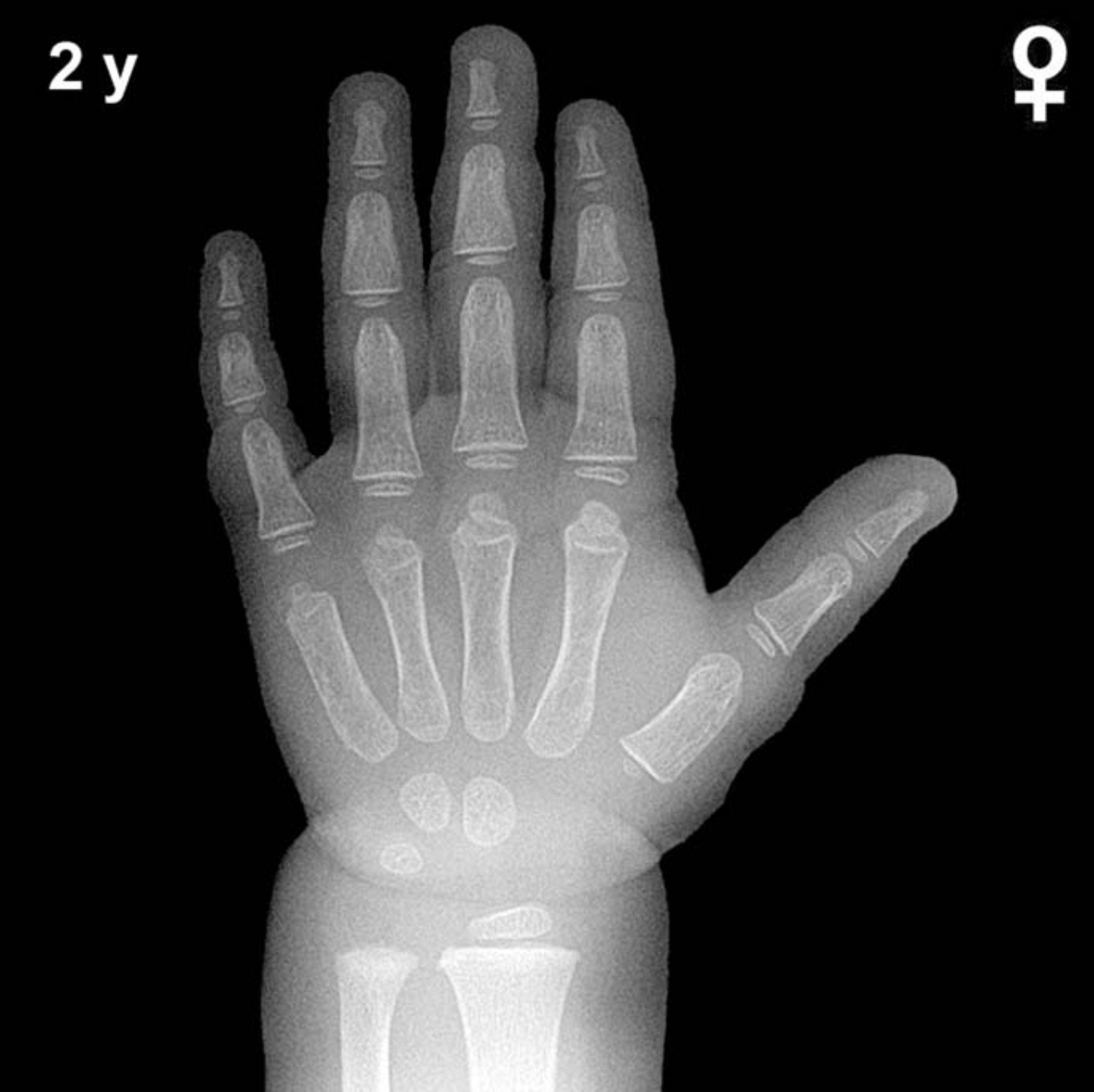
By 2 years of age in girls, several carpal and epiphyseal ossification centers are typically visible on a hand and wrist radiograph. The capitate and hamate are the earliest carpal bones to ossify, appearing around 3 and 6 months of age respectively, and are well established by this age. The triquetral ossification center typically emerges between 2 and 3 years in girls and may be just appearing or absent at exactly 2 years, making it an important landmark to note.
The distal radial epiphysis is generally present by approximately 1 year of age and should be clearly visible at 2 years. The lunate ossification center typically appears between 3 and 4 years in girls and is not yet expected at this age. Similarly, the scaphoid, trapezium, and trapezoid ossification centers are not anticipated until approximately 4–6 years. Epiphyses of the proximal and middle phalanges and metacarpals are typically present and developing by age 2, appearing as small, well-defined ossification centers at the bases of these bones.
- Capitate: Present (ossifies ~3 months)
- Hamate: Present (ossifies ~6 months)
- Distal radial epiphysis: Present (ossifies ~1 year)
- Triquetral: May be just appearing or absent at exactly 2 years
- Lunate, scaphoid, trapezium, trapezoid, pisiform: Not yet expected
Clinical Pearls
Skeletal maturation in girls is consistently ahead of boys by approximately 1–2 years throughout childhood, a well-recognized sex difference reflected in the Greulich-Pyle atlas. At 2 years, a bone age advance of 6–12 months may suggest conditions such as precocious puberty or excess androgen exposure, while a delay of similar magnitude may raise concern for growth hormone deficiency, hypothyroidism, or constitutional delay of growth and development. Turner syndrome should be considered in girls with persistently delayed bone age alongside other clinical features. A key pitfall at this age is over-reliance on the presence or absence of the triquetral alone, as its timing is highly variable in normal children; the overall pattern of ossification should guide interpretation rather than any single center. Reference: Greulich WW, Pyle SI. Radiographic Atlas of Skeletal Development of the Hand and Wrist. 2nd ed. Stanford University Press, 1959.