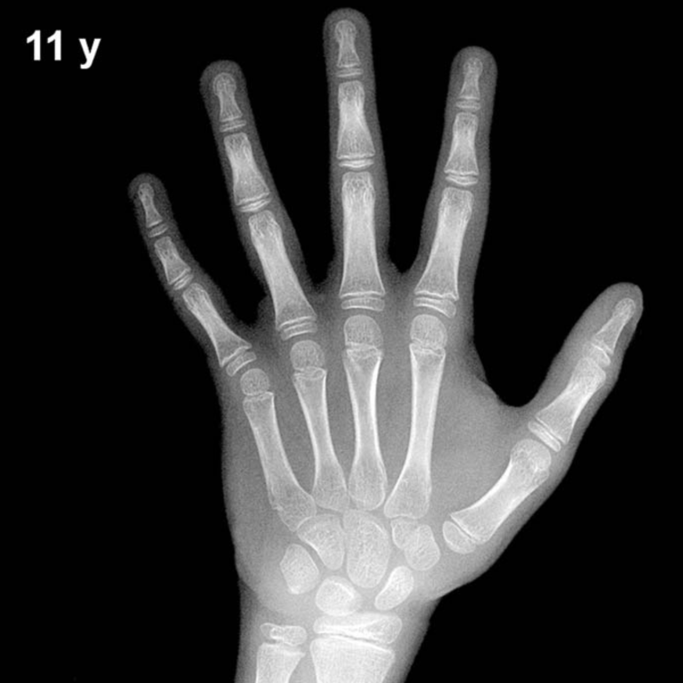
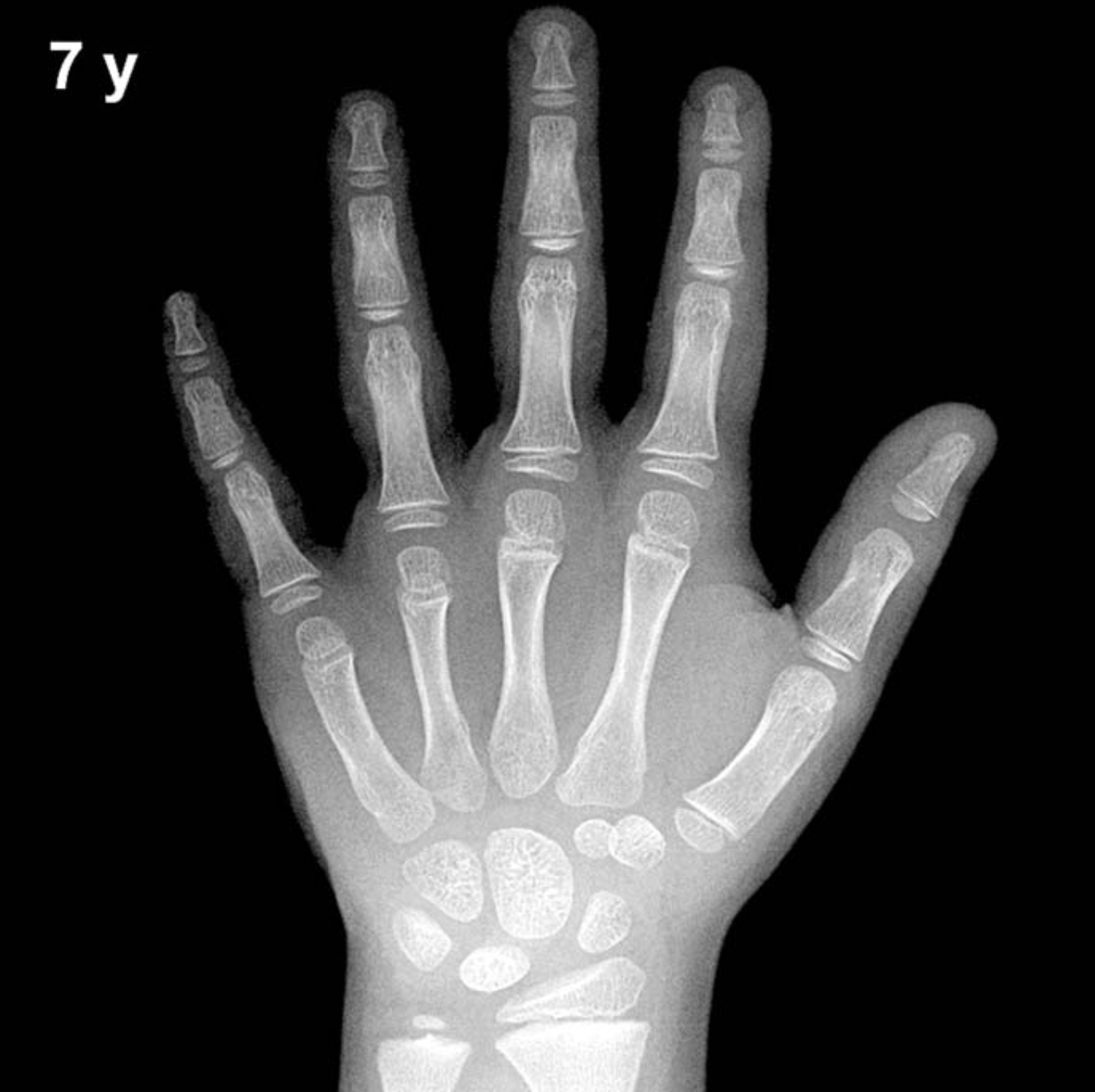
Bone Age in Boys Aged 7 Years — Greulich-Pyle Hand and Wrist X-Ray Reference
Bone age assessment using a left hand and wrist radiograph is a fundamental tool in pediatric radiology, allowing clinicians to estimate skeletal maturity independent of chronological age. The Greulich-Pyle (GP) method compares a child’s radiograph against standardized atlas plates derived from a mid-twentieth-century North American reference population. In 7-year-old boys, this assessment is particularly valuable in evaluating growth concerns, suspected endocrine disorders, and constitutional growth variants.
Expected Ossification Centers and Skeletal Findings
By 7 years of age in boys, all primary carpal ossification centers are typically present. The capitate and hamate are well-established (appearing around 3 and 6 months of life, respectively), and the triquetral, lunate, scaphoid, trapezium, and trapezoid are all expected to be visible by this age. The pisiform is generally not yet ossified in boys at 7 years, as it typically appears between approximately 11 and 14 years in males.
Epiphyseal development at this age includes a well-formed distal radial epiphysis (present from roughly 1 year of age) and a distal ulnar epiphysis that is typically visible, having appeared between approximately 5 and 7 years. The metacarpal and proximal, middle, and distal phalangeal epiphyses are present and show progressive broadening and maturation, though they remain clearly separated from their respective metaphyses at this age. The thumb sesamoid is not yet expected, as it typically appears in the peripubertal period.
- Capitate and hamate: well ossified
- Triquetral, lunate, scaphoid, trapezium, trapezoid: present
- Pisiform: absent (expected ~11–14 years in boys)
- Distal radial and ulnar epiphyses: present
- Metacarpal and phalangeal epiphyses: present, immature
- Thumb sesamoid: absent
Clinical Pearls
Skeletal maturation in girls is consistently ahead of boys, with a difference of approximately 1–2 years across mid-childhood. At 7 years, a normal variation of roughly ±1 year (approximately 1 standard deviation) is expected when comparing bone age to chronological age. A significantly advanced bone age in a 7-year-old boy may prompt evaluation for precocious puberty, congenital adrenal hyperplasia, or exogenous androgen exposure. Conversely, a delayed bone age may suggest growth hormone deficiency, hypothyroidism, or constitutional delay of growth and puberty, the latter being particularly common in boys.
A key interpretive pitfall is over-reliance on a single bone or center; the GP method requires a holistic comparison with the atlas plate. Population differences from the original reference cohort may also influence interpretation. Reference: Greulich WW, Pyle SI. Radiographic Atlas of Skeletal Development of the Hand and Wrist. 2nd ed. Stanford University Press, 1959.