Bone Age in Boys Aged 11 Years — Greulich-Pyle Hand and Wrist X-Ray Reference
Bone age assessment using a left-hand and wrist radiograph is a cornerstone of pediatric endocrine and growth evaluation. The Greulich-Pyle method compares a patient’s skeletal maturation against standardized atlas plates derived from a reference population. In boys aged 11 years, accurate bone age estimation informs workup for precocious puberty, growth hormone deficiency, hypothyroidism, and constitutional growth delay.
Expected Ossification Centers and Skeletal Findings
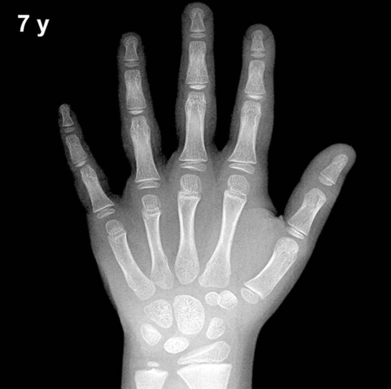
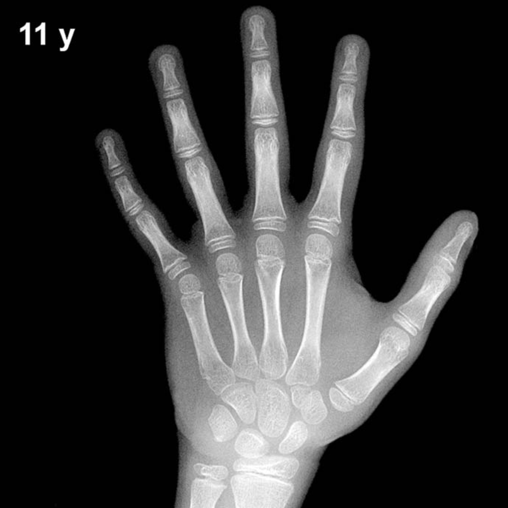
By 11 years in boys, all eight carpal bones are typically ossified. The capitate and hamate appear in early infancy (approximately 3 and 6 months, respectively), followed by the triquetral (2–3 years), lunate (3–4 years), scaphoid, trapezium, and trapezoid (4–6 years). The pisiform, which appears later in boys than in girls, is generally expected to be present or emerging by approximately 11–13 years in boys, and may be visible at this age in more advanced individuals.
Epiphyseal development at 11 years in boys is well advanced. The distal radial epiphysis (present by approximately 1 year) is now broad and well defined, and the distal ulnar epiphysis (typically appearing at 5–7 years) should be clearly visible. Metacarpal and phalangeal epiphyses are present at all levels, with progressive widening and capping of the growth plates reflecting ongoing pubertal maturation. The adductor sesamoid of the thumb typically appears in the peripubertal period; in boys it is often not yet present at 11 years but may emerge around 12–13 years in those with average or advanced skeletal maturity.
- Carpal bones: All eight expected to be ossified
- Pisiform: May be present or just emerging; appearance variable at this age
- Distal radius and ulna epiphyses: Well established and clearly visible
- Thumb sesamoid: Typically absent or just appearing; emergence signals peripubertal onset
Clinical Pearls
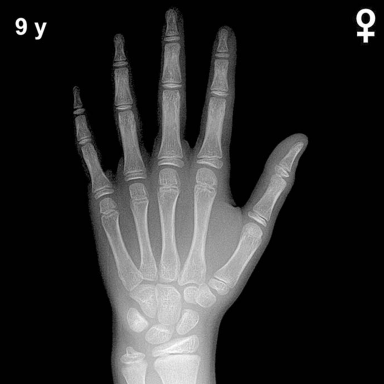
Girls are skeletally ahead of boys by approximately 1–2 years throughout childhood and adolescence, so an 11-year-old boy’s bone age atlas plate corresponds roughly to a 9–10-year-old girl. A bone age advanced by more than 2 standard deviations (roughly ≥2 years ahead) at this age should prompt evaluation for precocious puberty, congenital adrenal hyperplasia, or exogenous androgen exposure. Conversely, a significantly delayed bone age raises concern for growth hormone deficiency, hypothyroidism, or constitutional delay of growth and puberty. A key interpretive pitfall is over-reliance on a single skeletal indicator; the Greulich-Pyle method requires a global assessment of multiple ossification centers and epiphyseal morphology rather than any one landmark in isolation.
Reference: Greulich WW, Pyle SI. Radiographic Atlas of Skeletal Development of the Hand and Wrist. 2nd ed. Stanford University Press, 1959.