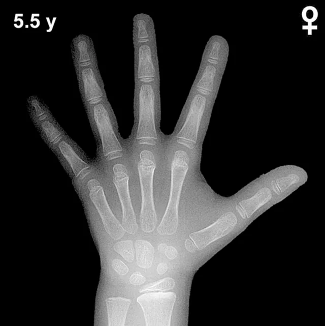
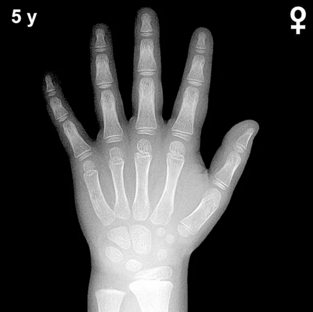
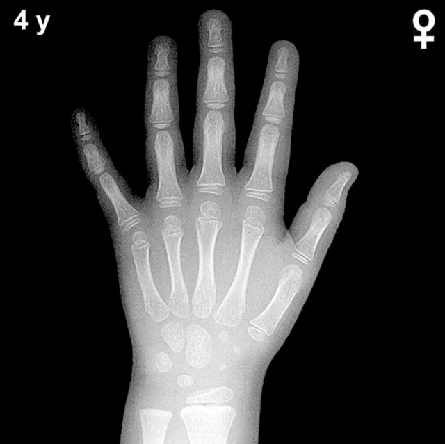
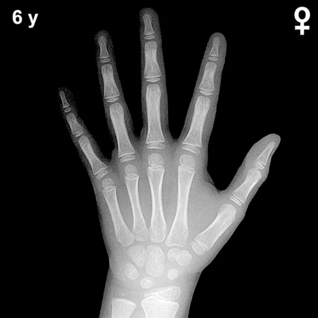
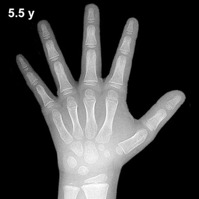
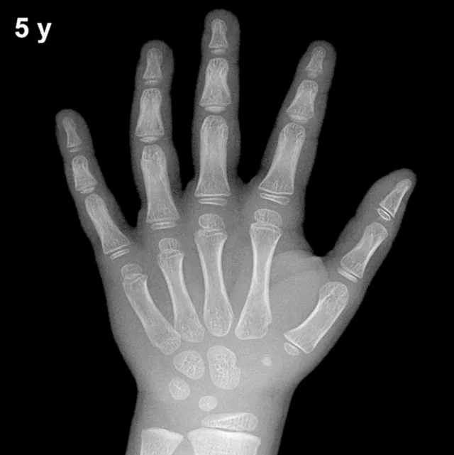
Bone Age in Girls Aged 3.5 Years — Greulich-Pyle Hand and Wrist X-Ray Reference
Bone age assessment using a posteroanterior hand and wrist radiograph is a standard method for evaluating skeletal maturity in children. The Greulich-Pyle (GP) atlas provides sex-specific reference standards by comparing a child's radiograph against atlas plates representing average skeletal development at defined ages. In girls aged 3.5 years, this assessment is clinically valuable in evaluating growth disorders, precocious puberty, hypothyroidism, and other endocrine conditions affecting skeletal maturation.
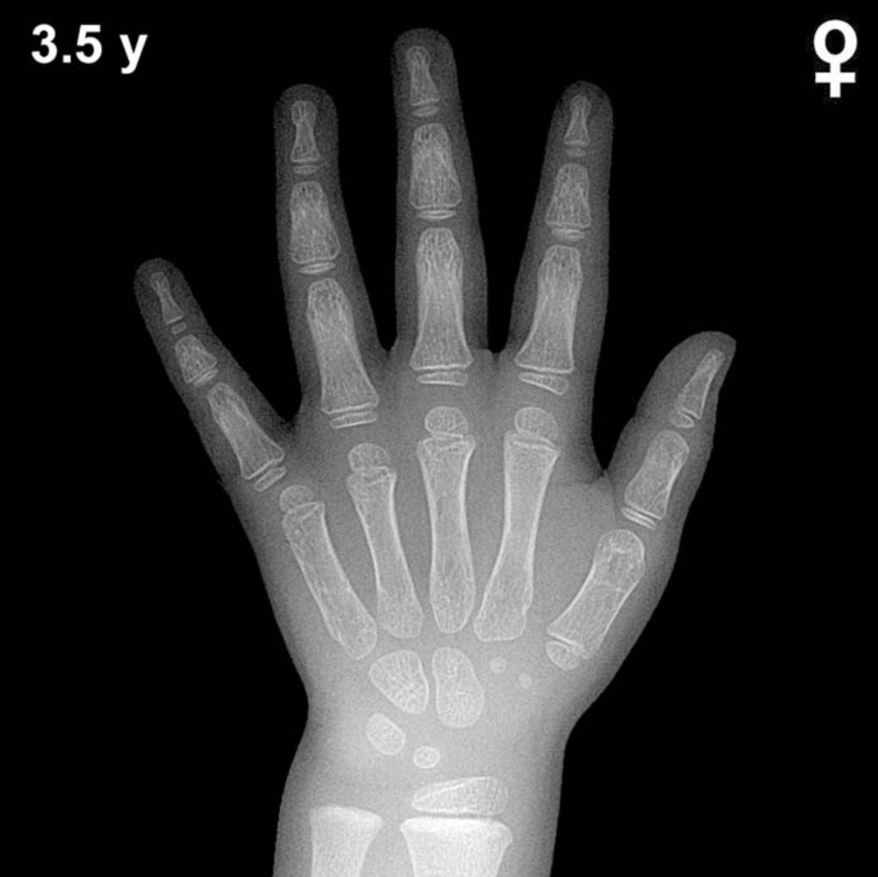
Expected Ossification Centers and Skeletal Findings
By 3.5 years of age in girls, several carpal ossification centers are typically well established. The capitate and hamate, the earliest carpal bones to ossify (appearing around 3 and 6 months respectively), should be clearly visible and well defined. The triquetral (which typically appears between 2–3 years) is generally present by this age, and the lunate, expected around 3–4 years, is usually visible or just emerging at 3.5 years in girls.
Epiphyseal development at this age includes a well-established distal radial epiphysis (typically appearing around 12 months) and developing epiphyses of the metacarpals and proximal, middle, and distal phalanges. The distal ulnar epiphysis is not yet expected at this age, as it typically appears between 5–7 years. The scaphoid, trapezium, and trapezoid are not yet reliably ossified at 3.5 years; these carpal bones generally appear between 4–6 years. The pisiform and the thumb sesamoid are absent, as expected, given their much later appearance.
- Present: Capitate, hamate, triquetral, lunate (emerging), distal radial epiphysis, phalangeal and metacarpal epiphyses
- Absent (expected): Distal ulnar epiphysis, scaphoid, trapezium, trapezoid, pisiform, thumb sesamoid
Clinical Pearls
Skeletal maturation in girls consistently leads that of boys by approximately 1–2 years during early childhood, a difference that widens further around puberty. At 3.5 years, a normal range of variability of roughly ±1 year is accepted. A bone age significantly advanced beyond 3.5 years in a girl this age may prompt evaluation for precocious puberty, congenital adrenal hyperplasia, or exogenous androgen exposure. Conversely, a notably delayed bone age raises concern for growth hormone deficiency, hypothyroidism, or constitutional delay of growth and development.
A key interpretive pitfall is over-reliance on a single carpal bone for staging; the GP method requires holistic assessment of multiple ossification centers and epiphyseal maturity together. Nutritional status and ethnicity may also influence skeletal maturation and should be considered when results fall outside expected ranges. Reference: Greulich WW, Pyle SI. Radiographic Atlas of Skeletal Development of the Hand and Wrist. 2nd ed. Stanford University Press, 1959.