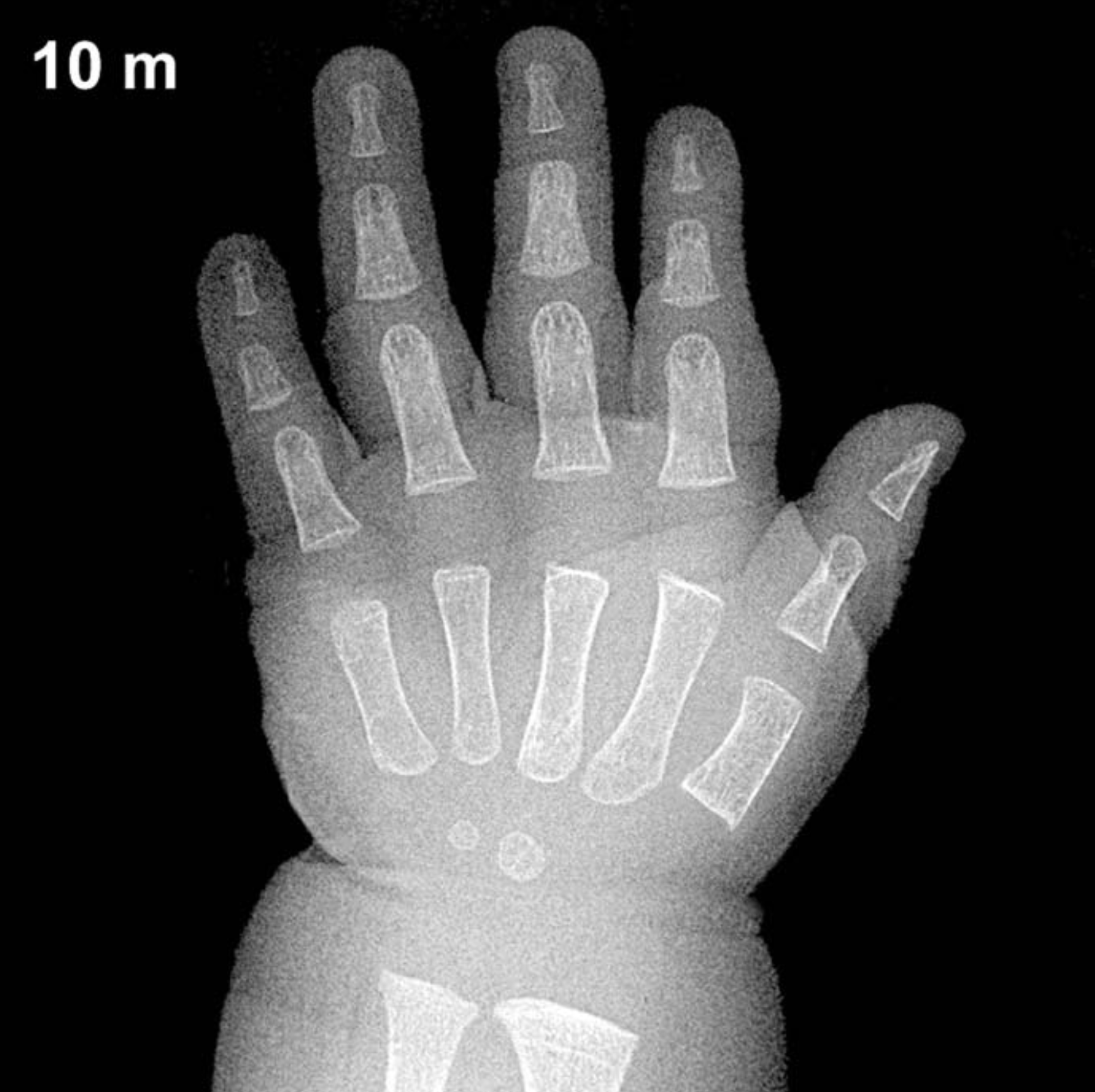
Bone Age in Boys Aged 10 Months — Greulich-Pyle Hand and Wrist X-Ray Reference
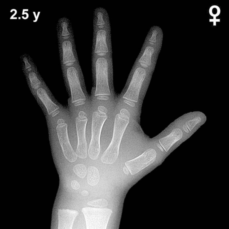
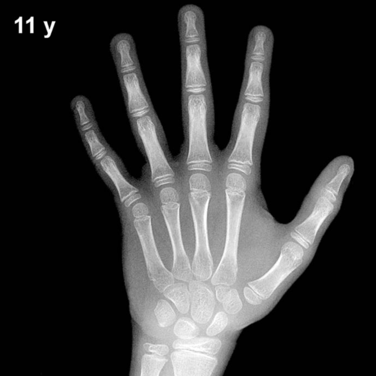
Bone age assessment using the Greulich-Pyle (GP) atlas compares a left-hand and wrist radiograph to sex-specific standard plates to estimate skeletal maturity. In infants, this evaluation is particularly useful in the workup of growth faltering, endocrine disorders such as congenital hypothyroidism, and — less commonly — forensic age estimation. Early identification of skeletal maturation delay or advancement in the first year of life can guide timely clinical intervention.
Expected Ossification Centers and Skeletal Findings
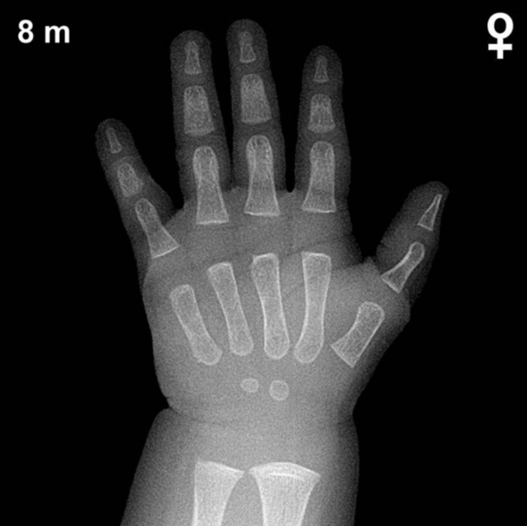
At 10 months of age in boys, the hand and wrist radiograph typically demonstrates a limited but important set of ossification centers. The capitate is the first carpal bone to ossify, usually appearing by approximately 3 months of age, and should be well established by 10 months. The hamate typically ossifies around 4–6 months and is also expected to be visible at this age. Together, the capitate and hamate are the only two carpal ossification centers reliably present in most boys at 10 months.
The remaining carpal bones — triquetral, lunate, scaphoid, trapezium, trapezoid, and pisiform — are not yet expected to be ossified at this age in boys. The distal radial epiphysis typically appears around 12 months in boys and may just be emerging or absent at 10 months; its presence or conspicuous absence should be noted. Metacarpal and phalangeal epiphyses are generally not yet ossified at this age. The distal ulnar epiphysis does not typically appear until 5–7 years of age.
- Capitate: present (ossifies ~3 months)
- Hamate: present (ossifies ~4–6 months)
- Distal radial epiphysis: absent or just emerging (~12 months in boys)
- All other carpal centers: absent at this age
Clinical Pearls
Skeletal maturation in girls is consistently ahead of boys throughout childhood; at 10 months, girls may already show early distal radial epiphyseal ossification, whereas boys typically lag slightly behind. The standard deviation for bone age at this age is approximately ±2–3 months, so minor variation from the expected pattern is normal. A bone age significantly delayed beyond this range should raise concern for congenital hypothyroidism, growth hormone deficiency, or other metabolic conditions, and warrants correlation with thyroid function tests and growth parameters. Conversely, markedly advanced skeletal maturation at this age — such as early appearance of multiple carpal centers — may suggest congenital adrenal hyperplasia or another source of androgen excess.
A key interpretive pitfall at this age is over-reliance on the number of ossification centers alone; positioning artifacts and radiographic exposure technique can obscure small, early-appearing centers or mimic their presence. Clinical correlation is always essential. Reference: Greulich WW, Pyle SI. Radiographic Atlas of Skeletal Development of the Hand and Wrist. 2nd ed. Stanford University Press, 1959.