Bone Age in Boys Aged 24 Months — Greulich-Pyle Hand and Wrist X-Ray Reference
Bone age assessment using a left-hand and wrist radiograph is a cornerstone of pediatric endocrine and growth evaluation, allowing clinicians to compare a child’s skeletal maturity against population norms. The Greulich-Pyle method matches the radiograph to standard atlas plates representing average skeletal development at specific ages. In boys at 24 months, this assessment helps identify early deviations suggestive of endocrine dysfunction, nutritional deficiency, or constitutional growth variants.
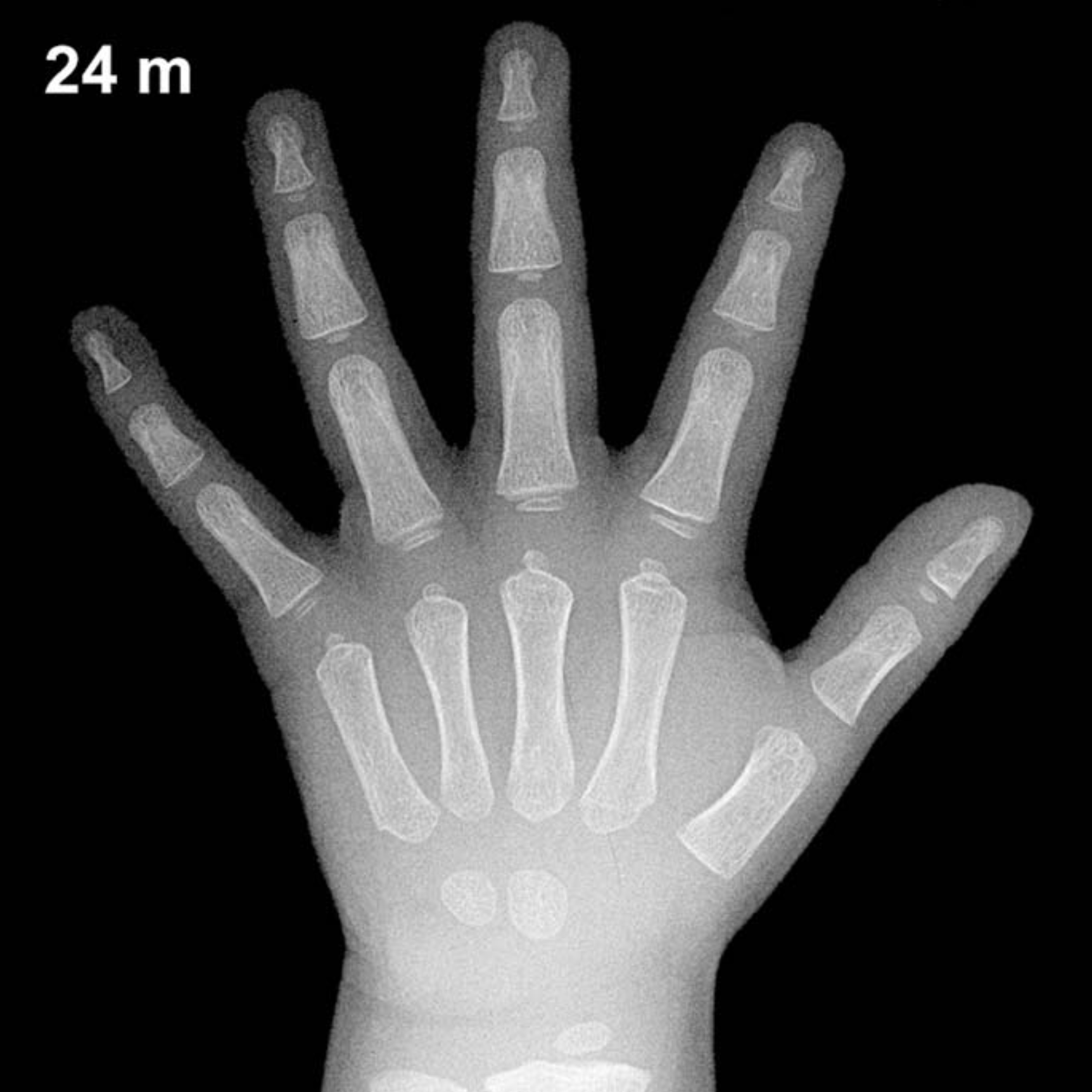
Expected Ossification Centers and Skeletal Findings
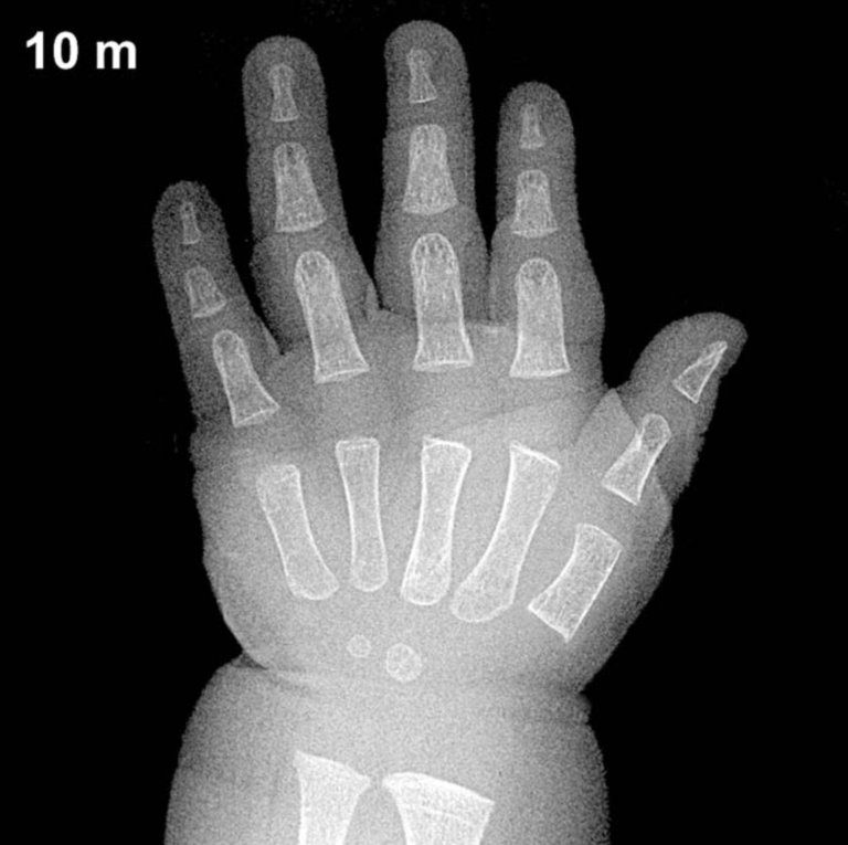
By 24 months in boys, several carpal ossification centers are typically visible on the hand and wrist radiograph. The capitate and hamate are the earliest carpal bones to ossify, appearing around 3 and 6 months respectively, and are well-established by this age. The triquetral ossification center typically emerges between 2 and 3 years in boys, so it may be just appearing or absent at exactly 24 months. The lunate generally follows between 3 and 4 years and is not expected at this age.
The distal radial epiphysis is typically present by approximately 12 months and should be clearly visible at 24 months. The distal ulnar epiphysis generally appears between 5 and 7 years and is not expected at this stage. Epiphyses of the proximal and middle phalanges, as well as the metacarpals, are typically ossifying and becoming more defined during this period. The scaphoid, trapezium, trapezoid, and pisiform ossification centers are not expected at 24 months in boys.
- Capitate: present (ossifies ~3 months)
- Hamate: present (ossifies ~6 months)
- Distal radial epiphysis: present (ossifies ~12 months)
- Triquetral: may be just emerging or absent at 24 months
- Lunate, scaphoid, trapezium, trapezoid, pisiform: not expected at this age
Clinical Pearls
Skeletal maturation in girls is known to run approximately 2–6 months ahead of boys during early childhood, a gap that widens further in adolescence. At 24 months, a normal range of bone age in boys spans roughly ±2 standard deviations around the mean, encompassing approximately 18 to 30 months of skeletal age. A bone age significantly advanced beyond chronological age may prompt evaluation for precocious puberty, congenital adrenal hyperplasia, or exogenous androgen exposure. Conversely, a notably delayed bone age raises consideration of growth hormone deficiency, hypothyroidism, or constitutional delay of growth and puberty.
A key interpretive pitfall at this age is over-reliance on a single ossification center: the triquetral, in particular, shows wide physiological variability in its timing of appearance. Clinical correlation with growth velocity, auxological data, and biochemical investigations remains essential. Reference: Greulich WW, Pyle SI. Radiographic Atlas of Skeletal Development of the Hand and Wrist. 2nd ed. Stanford University Press, 1959.