Bone Age in Boys Aged 8 Years — Greulich-Pyle Hand and Wrist X-Ray Reference
Bone age assessment using a left-hand and wrist radiograph is a fundamental tool in pediatric radiology for evaluating skeletal maturation relative to chronological age. The Greulich-Pyle method compares a child’s radiograph against standardized atlas plates to assign a skeletal age, guiding clinical decisions in growth disorders, endocrine evaluation, and, where necessary, forensic age estimation. In 8-year-old boys, the hand and wrist radiograph reflects a mid-childhood pattern of progressive epiphyseal and carpal development that is well characterized in the atlas.

Expected Ossification Centers and Skeletal Findings
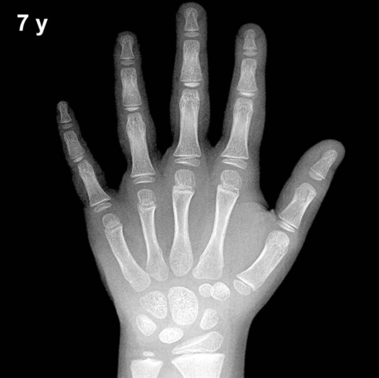
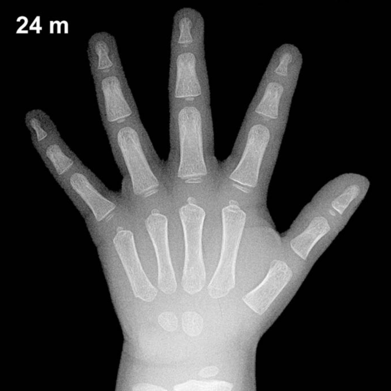
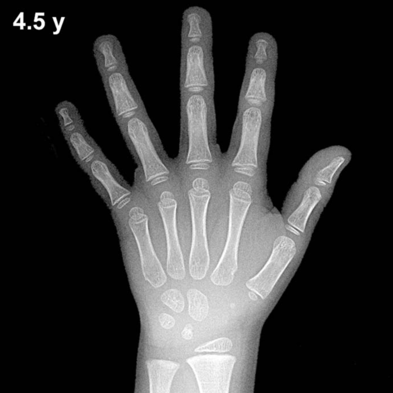
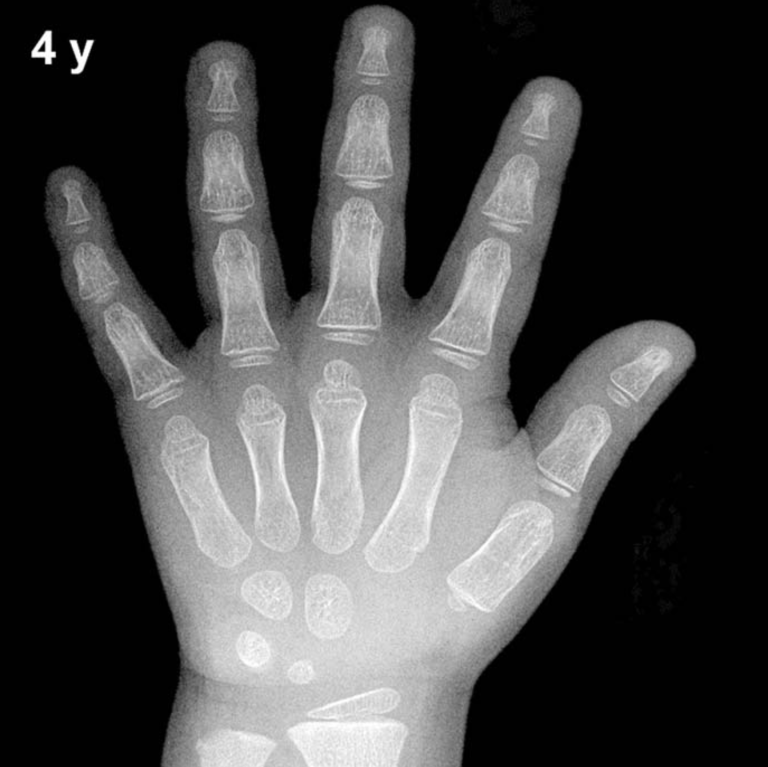
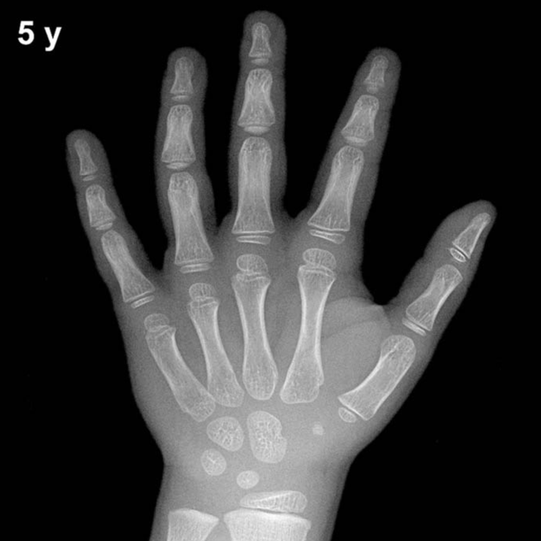
By 8 years of age in boys, all eight carpal bones are typically ossified and visible on the radiograph. The capitate and hamate appear earliest in infancy (approximately 3 and 6 months, respectively), followed by the triquetral (around 2–3 years), lunate (around 3–4 years), scaphoid, trapezium, and trapezoid (approximately 4–6 years). By age 8, these centers should be well established and show progressive growth and definition in size and contour.
The distal radial epiphysis, which typically appears around 1 year of age, is now well developed and broad at this stage. The distal ulnar epiphysis, which generally appears between 5 and 7 years, should be clearly visible and beginning to widen. Epiphyses of the metacarpals and phalanges are present and showing increasing maturation, with the proximal phalangeal epiphyses particularly prominent.
- Capitate and hamate: Long ossified; well-defined and of substantial size
- Triquetral, lunate, scaphoid, trapezium, trapezoid: All present and progressively enlarging
- Distal radial and ulnar epiphyses: Well visualized; distal ulnar epiphysis established
- Metacarpal and phalangeal epiphyses: Present throughout; increasing definition
- Pisiform: Not yet expected; typically appears around 9–12 years in girls and 11–14 years in boys
- Thumb sesamoid: Not yet expected; typically appears peripu bertally
Clinical Pearls
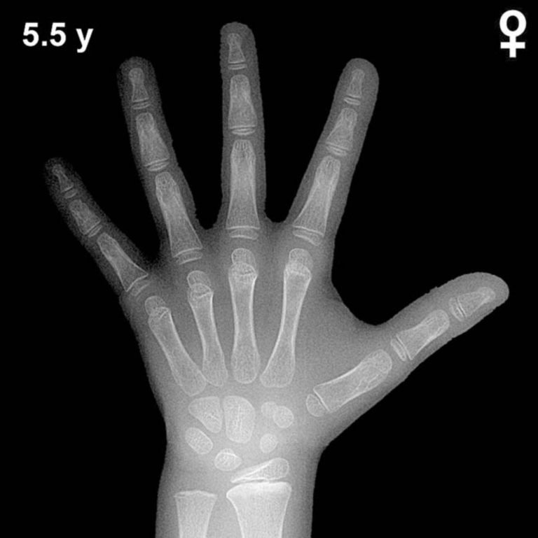
Skeletal maturation in girls is consistently ahead of boys by approximately 1–2 years across childhood, a difference well documented in the Greulich-Pyle atlas. At chronological age 8, a bone age within roughly ±2 standard deviations (approximately ±1–1.5 years) is generally considered within normal limits, though population-specific variation should be acknowledged. An advanced bone age at this age may prompt evaluation for precocious puberty, congenital adrenal hyperplasia, or exogenous androgen exposure. A delayed bone age raises concern for growth hormone deficiency, hypothyroidism, constitutional delay of growth and puberty, or chronic systemic illness.
A key interpretive pitfall is over-reliance on a single skeletal indicator; bone age should be assigned by overall pattern matching to the atlas plate rather than any isolated center. Nutritional status and ethnicity may also influence skeletal maturation, and the original Greulich-Pyle sample was derived from a mid-20th-century North American population of European descent. Reference: Greulich WW, Pyle SI. Radiographic Atlas of Skeletal Development of the Hand and Wrist. 2nd ed. Stanford University Press, 1959.