Bone Age in Girls Aged 13 Years — Greulich-Pyle Hand and Wrist X-Ray Reference
Bone age assessment using the Greulich-Pyle atlas involves comparing a left-hand and wrist radiograph to standardized reference plates to estimate skeletal maturity. In girls aged 13 years, this evaluation is particularly relevant in the context of pubertal timing, growth disorders, and endocrine conditions such as precocious puberty or growth hormone deficiency. Accurate bone age determination at this stage guides clinical decisions around growth potential and treatment planning.
Expected Ossification Centers and Skeletal Findings
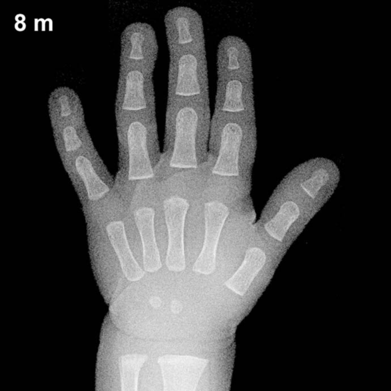
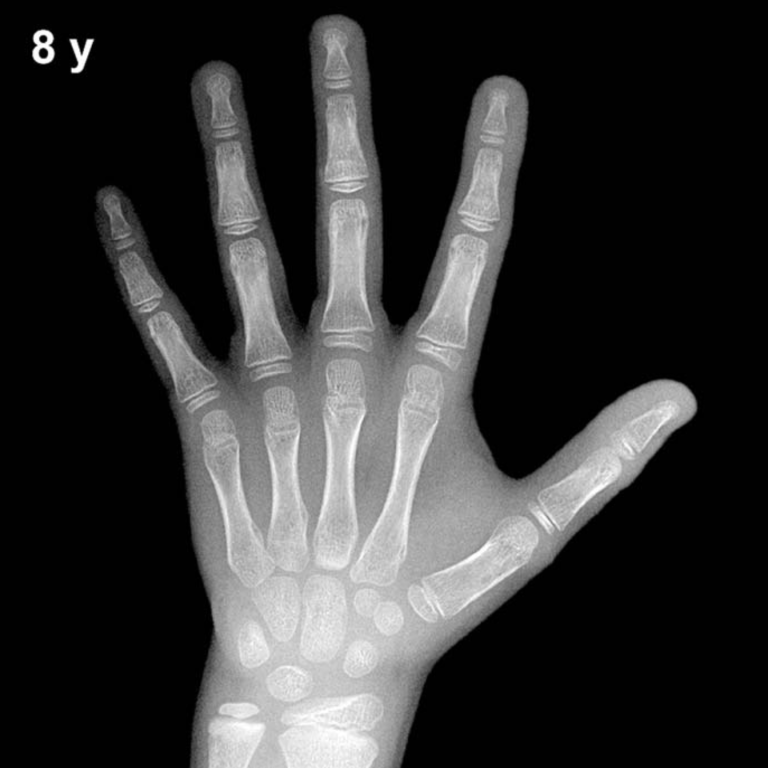
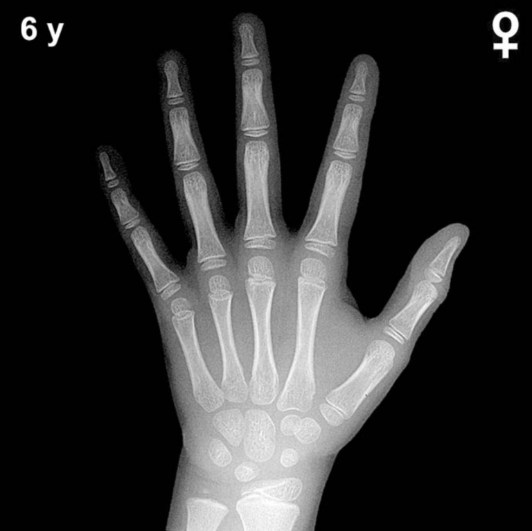
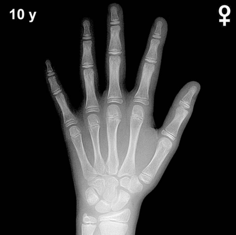
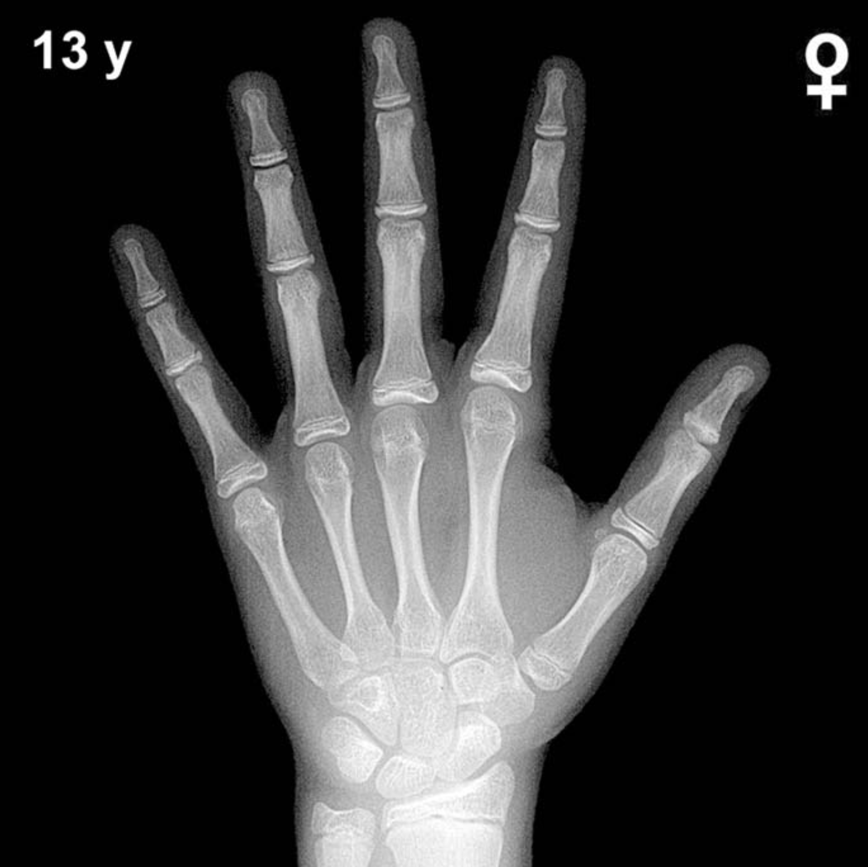
By 13 years in girls, all primary carpal ossification centers are well established. The capitate (present from approximately 3 months), hamate (~6 months), triquetral (~2–3 years), lunate (~3–4 years), scaphoid, trapezium, and trapezoid (~4–6 years) should all be fully visible and maturing. The pisiform, which typically appears between 9–12 years in girls, is expected to be present and well ossified by this age.
Epiphyseal maturation is advanced at 13 years in girls. The distal radial epiphysis, present since approximately 1 year of age, typically shows significant widening and capping of the metaphysis. The distal ulnar epiphysis, appearing around 5–7 years, should be well developed. A hallmark finding at this age is the sesamoid of the thumb (adductor pollicis), which appears in the peripubertal period and is typically present by 13 years in girls, often serving as a useful maturation landmark.
- All eight carpal bones ossified and maturing
- Pisiform present and well ossified
- Thumb sesamoid (adductor pollicis) typically visible
- Distal radial and ulnar epiphyses broad, with progressive physeal narrowing
- Phalangeal epiphyses showing advanced capping; some may approach fusion in early-maturing girls
Clinical Pearls
At 13 years, girls’ skeletal maturity is typically 1.5–2 years ahead of boys of the same chronological age, reflecting the well-documented sex difference in skeletal maturation. Normal variability is approximately ±1 standard deviation of roughly 12–15 months at this age, so bone age between approximately 11.5 and 14.5 years may fall within the expected range. A bone age significantly advanced beyond 14.5 years should prompt consideration of precocious puberty or exogenous androgen/estrogen exposure, while a bone age below 11.5 years may suggest constitutional delay of growth and puberty, hypothyroidism, or growth hormone deficiency.
A key interpretive pitfall at this age is overestimating skeletal maturity based solely on epiphyseal width without assessing physeal fusion status; early fusion at the phalanges can indicate markedly advanced maturation and potential compromise of final adult height. Reference: Greulich WW, Pyle SI. Radiographic Atlas of Skeletal Development of the Hand and Wrist. 2nd ed. Stanford University Press, 1959.