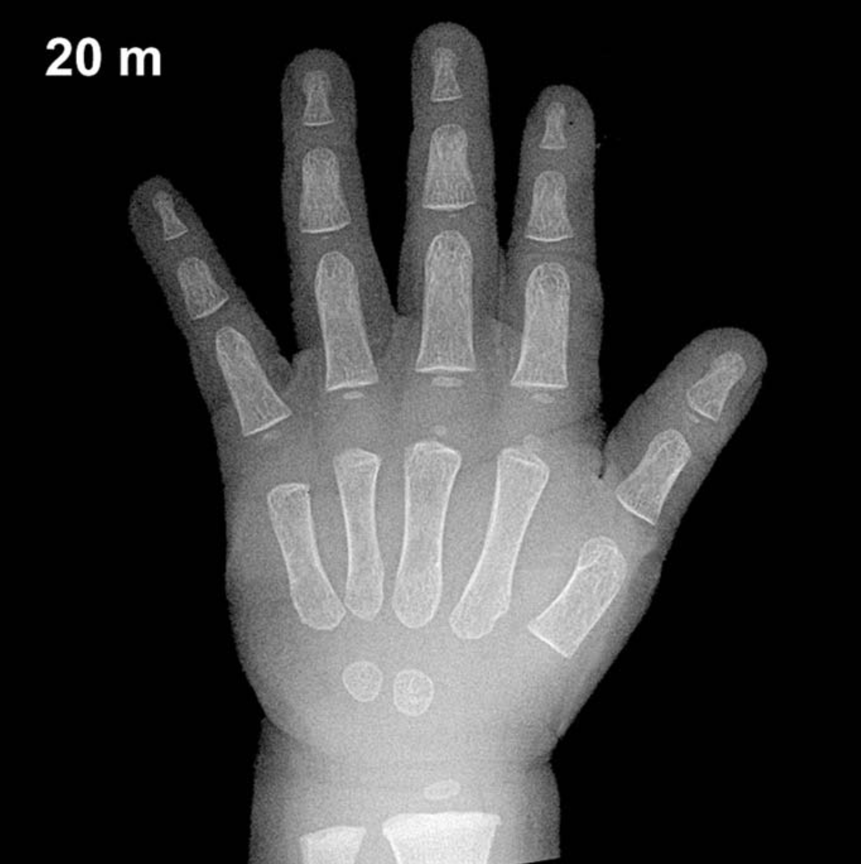
Bone Age in Boys Aged 20 Months — Greulich-Pyle Hand and Wrist X-Ray Reference
Bone age assessment using the Greulich-Pyle (GP) atlas involves comparing a left-hand and wrist radiograph to standardized reference plates to estimate skeletal maturity. In toddlers, this evaluation is particularly valuable when investigating growth faltering, suspected endocrine disorders, or when chronological age is uncertain in forensic or adoption contexts. Early identification of skeletal advancement or delay can guide timely workup and intervention.
Expected Ossification Centers and Skeletal Findings
At 20 months in boys, the Greulich-Pyle atlas anticipates a relatively limited but consistent set of ossification centers in the hand and wrist. The capitate and hamate are the earliest carpal bones to appear, typically ossified by 3–6 months of age and therefore well established by 20 months. The triquetral ossification center, which typically emerges between approximately 2 and 3 years in boys, may be absent or just beginning to appear at this age; its presence would suggest skeletal advancement relative to chronological age.
The distal radial epiphysis is generally visible by around 12 months and should be clearly present at 20 months, appearing as a thin, rounded ossification center. The lunate, scaphoid, trapezium, and trapezoid are not expected to be ossified at this age in boys, as these typically appear between 3 and 6 years. The pisiform and distal ulnar epiphysis are likewise absent at this stage. Epiphyseal ossification centers of the metacarpals and proximal phalanges may be visible as small, flat ossific nuclei, consistent with early childhood maturation.
- Present: Capitate, hamate, distal radial epiphysis
- Emerging or absent: Triquetral (may just appear near 2 years)
- Not yet expected: Lunate, scaphoid, trapezium, trapezoid, pisiform, distal ulnar epiphysis
Clinical Pearls
Skeletal maturation in girls is typically 2–6 months ahead of boys at this early age, so the GP standard plates for a 20-month-old boy reflect a slightly less mature pattern than those for a girl of the same chronological age. A bone age advanced by more than 2 standard deviations should prompt consideration of precocious puberty, congenital adrenal hyperplasia, or exogenous androgen exposure. Conversely, significant bone age delay may suggest growth hormone deficiency, hypothyroidism, or constitutional delay of growth and puberty. A key interpretive pitfall at this age is over-reliance on a single ossification center: the presence or absence of the triquetral alone should not drive clinical decision-making without correlating with the overall skeletal maturity pattern and clinical context. Reference: Greulich WW, Pyle SI. Radiographic Atlas of Skeletal Development of the Hand and Wrist. 2nd ed. Stanford University Press, 1959.