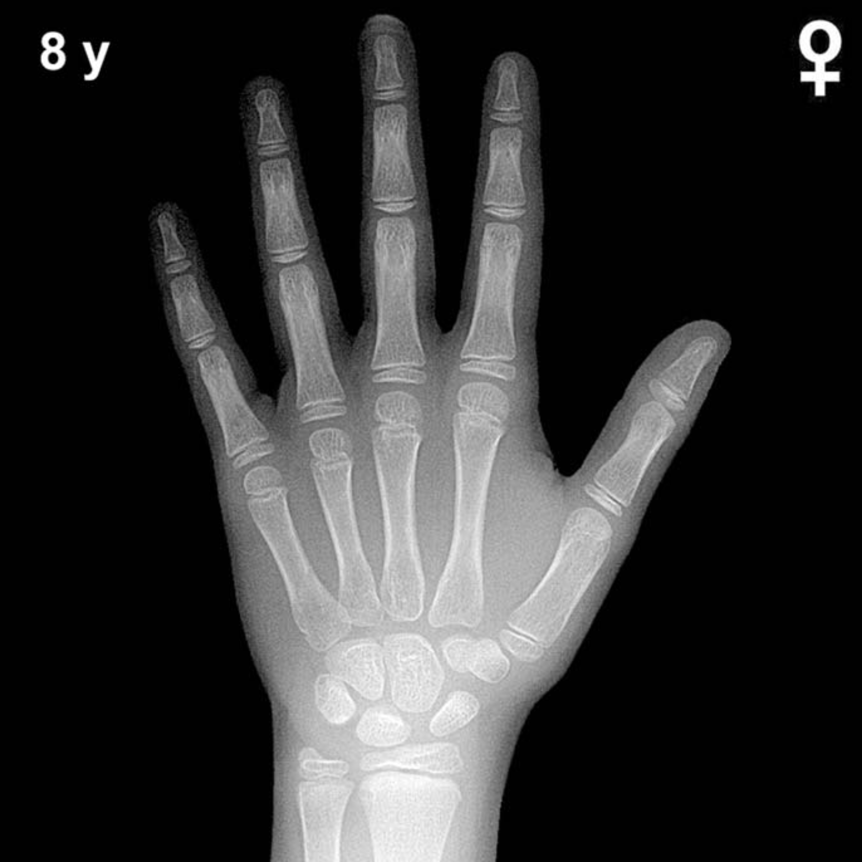
Bone Age in Girls Aged 8 Years — Greulich-Pyle Hand and Wrist X-Ray Reference
Bone age assessment using a left hand and wrist radiograph is a standard tool for evaluating skeletal maturity in children. The Greulich-Pyle method compares a patient’s radiograph against sex-specific reference plates to estimate skeletal age. In 8-year-old girls, this assessment is commonly performed during workup for growth disturbances, early or delayed puberty, and endocrine conditions such as thyroid or growth hormone disorders.
Expected Ossification Centers and Skeletal Findings
By 8 years of age in girls, all eight carpal bones are typically ossified. The capitate and hamate appear in early infancy, while the triquetral, lunate, scaphoid, trapezium, and trapezoid generally ossify progressively through the preschool years. The pisiform, which ossifies later than other carpals, is typically visible by approximately 8–9 years in girls and may be just appearing or well established at this age.
The epiphyses of the distal radius and distal ulna are well developed by this age. Epiphyseal maturation is evident across the metacarpals and phalanges, with progressive capping of the metaphyses. The epiphyses of the proximal, middle, and distal phalanges should show increasing breadth relative to their respective diaphyses. The adductor sesamoid of the thumb has not typically appeared at 8 years and its presence would suggest advanced skeletal maturity or early pubertal onset.
- Carpal bones: All eight ossification centers typically present, including a visible pisiform.
- Distal radius and ulna: Epiphyses well established and broadening.
- Metacarpal and phalangeal epiphyses: Progressively widening, consistent with mid-childhood growth.
- Thumb sesamoid: Typically absent at this age in girls.
Clinical Pearls
Girls’ skeletal maturation is consistently ahead of boys’ by approximately 1–2 years at this chronological age. A bone age more than 2 standard deviations above the mean (roughly ≥10 years skeletal age) should prompt consideration of precocious puberty, congenital adrenal hyperplasia, or exogenous androgen/estrogen exposure. Conversely, a bone age significantly below the mean may suggest growth hormone deficiency, hypothyroidism, or constitutional delay of growth and puberty. Bone age delay in a short-statured girl should also raise consideration of Turner syndrome, particularly if other clinical features are present.
A key interpretive pitfall is over-relying on a single carpal or epiphyseal landmark rather than assessing the overall pattern of skeletal maturation across all visible structures. Individual bone variability is normal, and the Greulich-Pyle method is best applied as a gestalt comparison to the atlas plates rather than a checklist of isolated findings. Reference: Greulich WW, Pyle SI. Radiographic Atlas of Skeletal Development of the Hand and Wrist. 2nd ed. Stanford University Press, 1959.