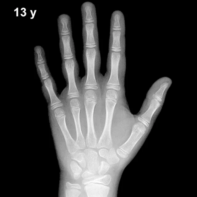
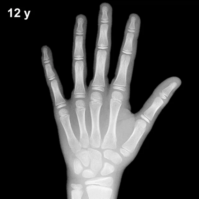
Bone Age in Boys Aged 15 Years — Greulich-Pyle Hand and Wrist X-Ray Reference
Bone age assessment using the Greulich-Pyle (GP) atlas compares a left-hand-and-wrist radiograph against standardized reference plates to estimate skeletal maturity. In boys aged 15 years, this evaluation is particularly relevant for investigating pubertal timing, growth hormone deficiency, constitutional delay of growth and puberty, and hypothyroidism. Accurate bone age determination helps clinicians predict adult height potential and guide endocrine management decisions.
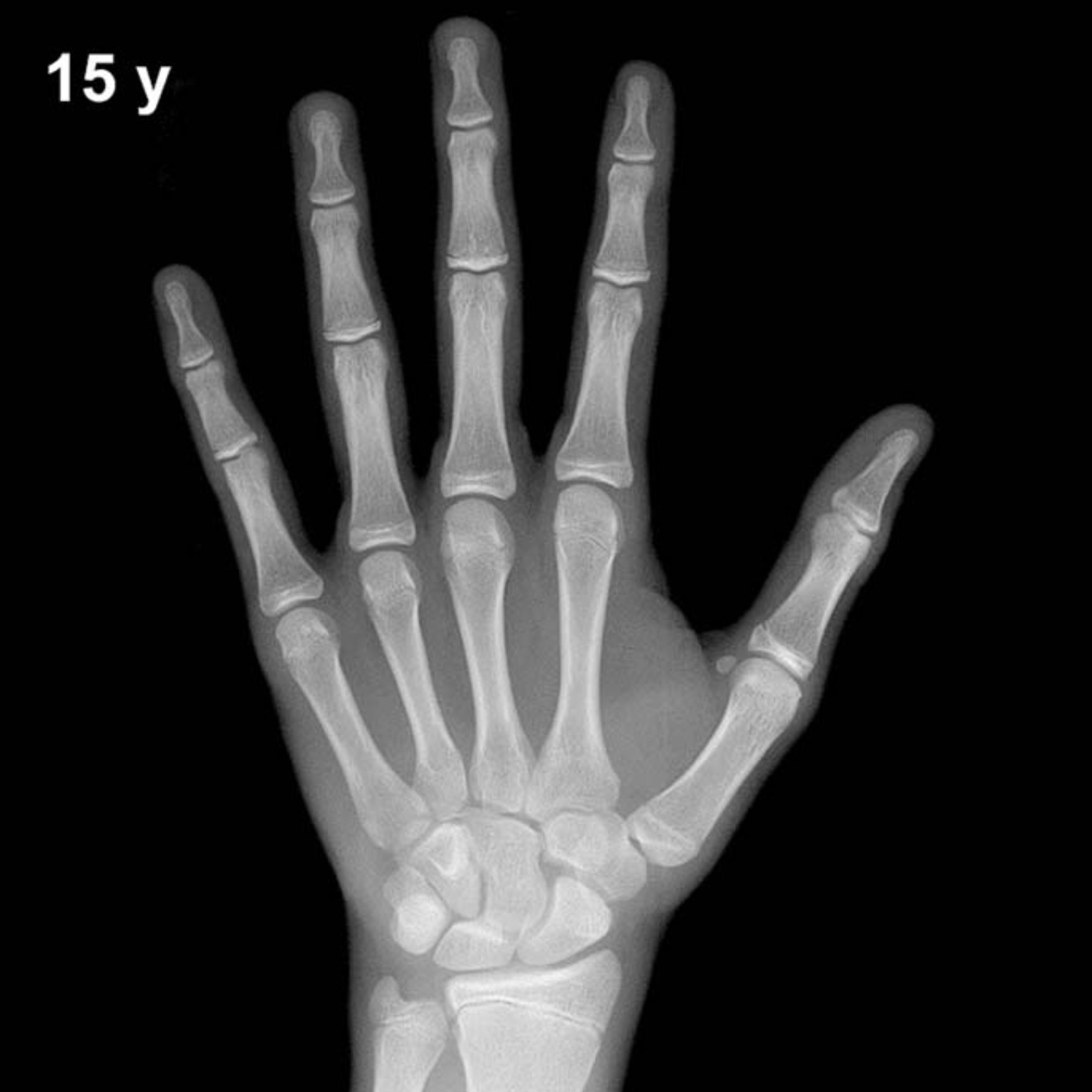
Expected Ossification Centers and Skeletal Findings
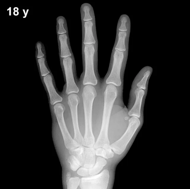
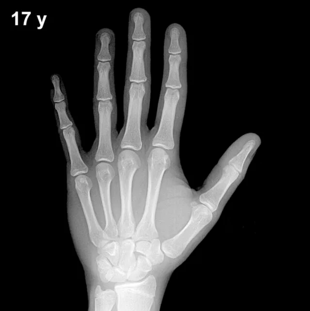
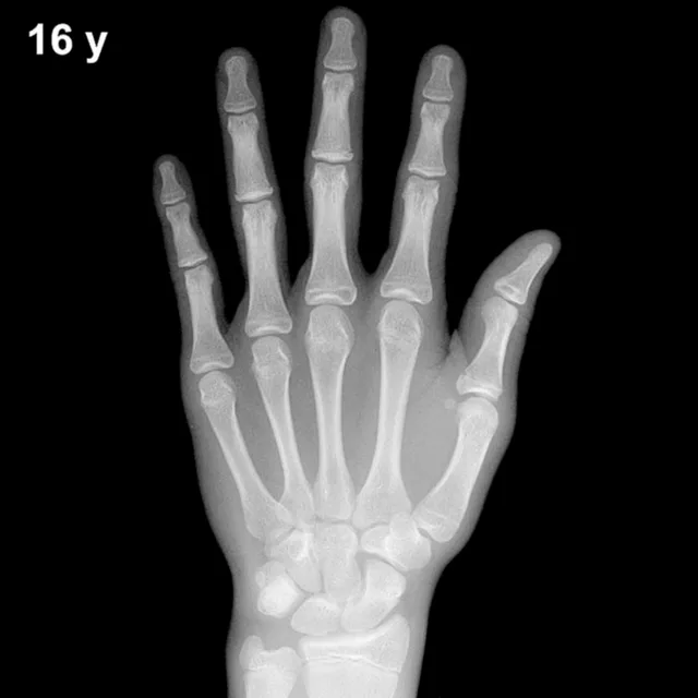
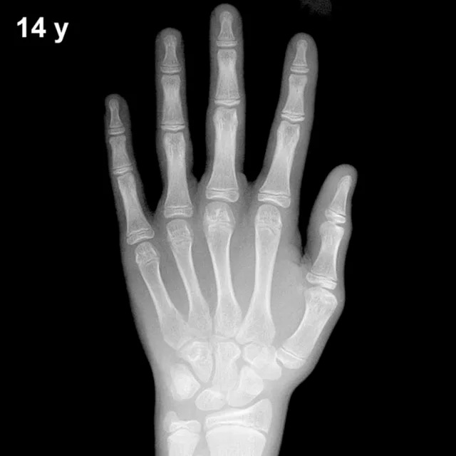
By 15 years in boys, all primary carpal ossification centers — capitate, hamate, triquetral, lunate, scaphoid, trapezium, and trapezoid — are well established and typically show mature, well-corticated morphology. The pisiform, which typically appears between approximately 11 and 14 years in boys, should be present by this age. All eight carpal bones are expected to be ossified and approaching adult configuration.
Epiphyseal development is advanced at 15 years. The distal radial and ulnar epiphyses are well formed, with the distal radial epiphysis typically spanning the full width of the metaphysis. Epiphyseal fusion in boys begins in earnest around this age: the proximal phalanges and middle phalanges of the fingers may show early or partial fusion at their bases in boys at the standard 15-year plate, while the distal phalanges may be further along in fusion. The adductor sesamoid of the thumb, a reliable peripubertal marker, is expected to be present and well ossified by this stage.
- Carpals: All eight centers present; mature morphology expected.
- Distal radius/ulna epiphyses: Well developed; radial epiphysis full-width.
- Phalangeal epiphyses: Cap-shaped or partially fusing, particularly in distal and proximal phalanges.
- Thumb sesamoid: Present and ossified.
- Pisiform: Should be visible by this age in boys.
Clinical Pearls
At 15 years, the standard deviation of bone age in boys is approximately ±1 year, meaning a skeletal age of 13–17 years may still fall within the broad population range. Girls' skeletal maturation is typically 1.5–2 years ahead of boys at equivalent chronological ages, which is an important consideration when interpreting population-based data. A bone age significantly advanced beyond 15 years in a 15-year-old boy raises concern for androgen excess, including congenital adrenal hyperplasia or an androgen-secreting tumor. Conversely, a bone age notably delayed (e.g., ≤13 years) warrants evaluation for constitutional delay of growth and puberty, growth hormone deficiency, or hypothyroidism. A key interpretive pitfall at this age is over-reliance on a single epiphyseal fusion event; fusion sequence shows inter-individual variability, and the overall pattern across multiple bones should guide assessment rather than any isolated finding. Reference: Greulich WW, Pyle SI. Radiographic Atlas of Skeletal Development of the Hand and Wrist. 2nd ed. Stanford University Press, 1959.