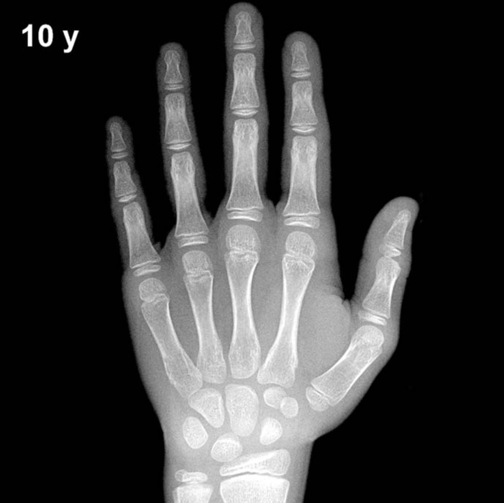
Bone Age in Boys Aged 10 Years — Greulich-Pyle Hand and Wrist X-Ray Reference
Bone age assessment using a left-hand and wrist radiograph is a cornerstone technique for evaluating skeletal maturity in children. The Greulich-Pyle (GP) method compares a child’s radiograph against standardized atlas plates to estimate biological age, which may differ from chronological age. Clinically, this evaluation is essential in the workup of growth disorders, precocious or delayed puberty, endocrine conditions, and medicolegal age estimation.
Expected Ossification Centers and Skeletal Findings
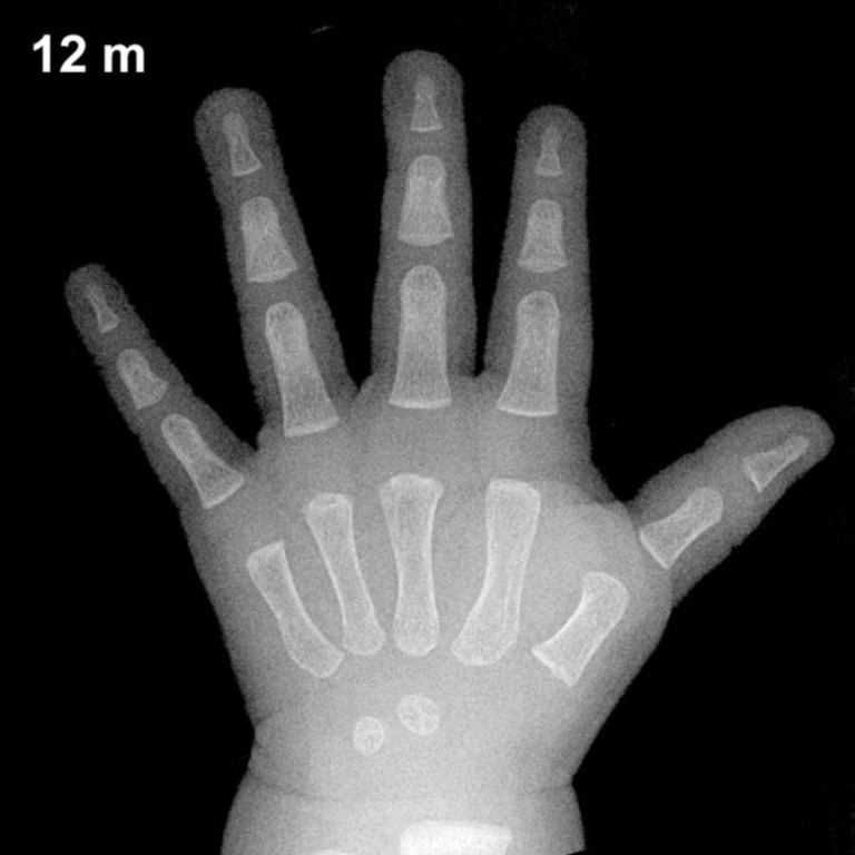
By 10 years of age in boys, all eight carpal bones are typically ossified and well established. The capitate and hamate appear in early infancy, followed by the triquetral (approximately 2–3 years), lunate (approximately 3–4 years), scaphoid, trapezium, and trapezoid (approximately 4–6 years). By age 10, these carpal centers should demonstrate increasing size and definition, with progressive maturation of articular margins.
The distal radial epiphysis is well ossified and broadening, typically showing increased density and a more squared configuration. The distal ulnar epiphysis, which appears around 5–7 years, should be clearly visible and growing. Epiphyses of the metacarpals and phalanges are present and enlarging, with the proximal phalangeal epiphyses showing characteristic widening. The pisiform, which typically ossifies between approximately 11–14 years in boys, is generally not yet visible at this age, and its absence is an expected finding.
- Capitate & hamate: Present and well ossified
- Triquetral, lunate, scaphoid, trapezium, trapezoid: All present; increasing maturity
- Pisiform: Typically absent at 10 years in boys
- Distal radial epiphysis: Well formed, broadening
- Distal ulnar epiphysis: Present and developing
- Thumb sesamoid: Not yet expected; typically appears peripubertally
Clinical Pearls
At age 10, boys’ skeletal maturity typically lags behind girls of the same chronological age by approximately 1–2 years, reflecting the well-documented sex difference in skeletal maturation. A bone age advanced by more than 2 standard deviations should prompt consideration of precocious puberty, congenital adrenal hyperplasia, or hyperthyroidism. Conversely, a delayed bone age may suggest growth hormone deficiency, hypothyroidism, or constitutional delay of growth and puberty. Turner syndrome is not applicable here but remains relevant in girls with delayed bone age.
A key interpretive pitfall is over-reliance on a single skeletal region; the GP method requires global assessment across multiple bones rather than any single epiphysis or carpal center. Population-based variation is significant, with normal ranges spanning approximately ±1–2 years at this age. Reference: Greulich WW, Pyle SI. Radiographic Atlas of Skeletal Development of the Hand and Wrist. 2nd ed. Stanford University Press, 1959.