Bone Age in Boys Aged 9 Years — Greulich-Pyle Hand and Wrist X-Ray Reference
Bone age assessment using a left-hand and wrist radiograph is a cornerstone of pediatric endocrine and growth evaluation. The Greulich-Pyle method compares a child’s skeletal maturation against sex-specific standard atlas plates derived from a mid-twentieth-century North American reference population. In 9-year-old boys, this assessment is particularly relevant in evaluating growth hormone deficiency, precocious puberty, constitutional delay, and hypothyroidism.
Expected Ossification Centers and Skeletal Findings
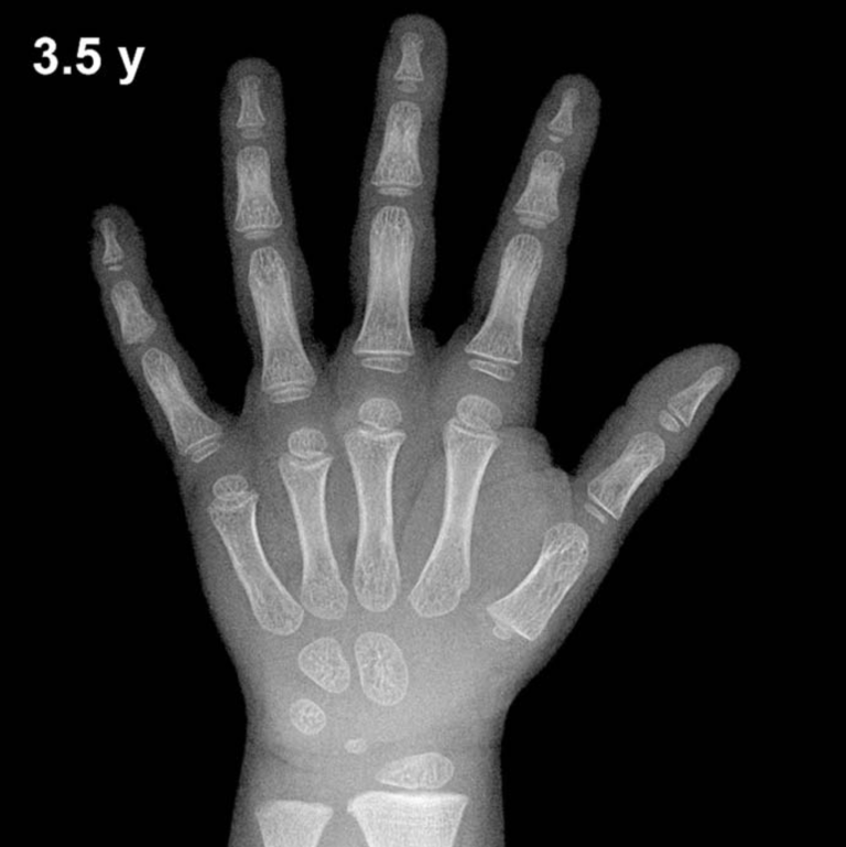
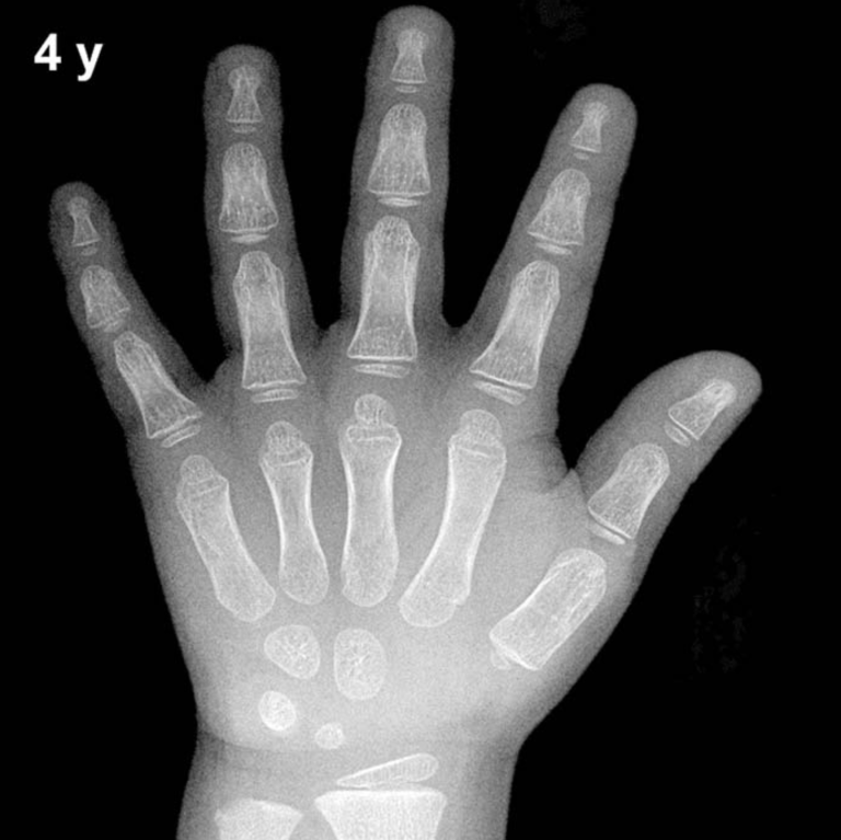
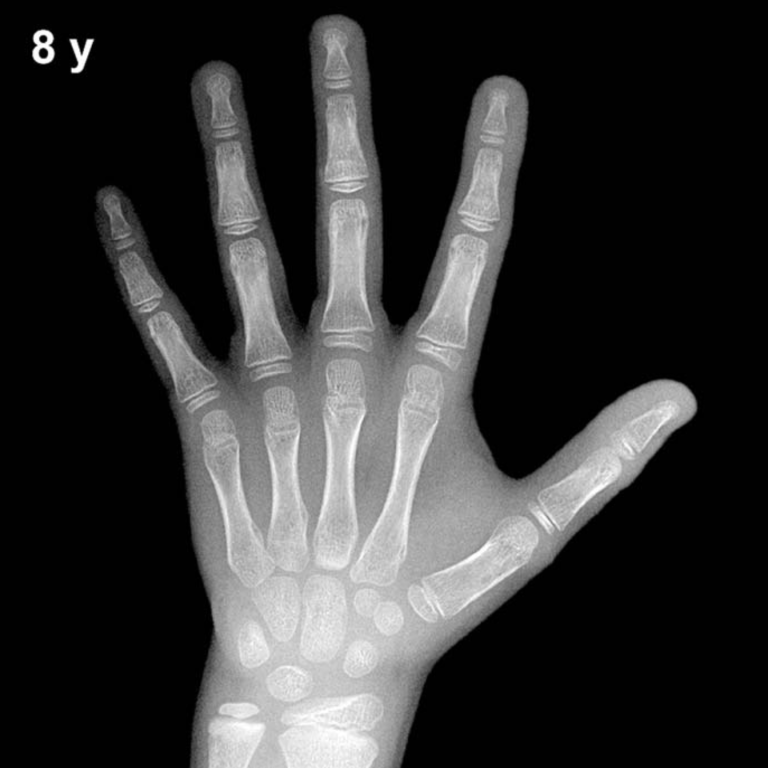
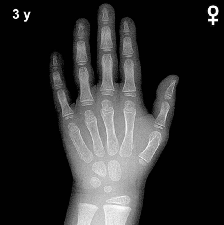
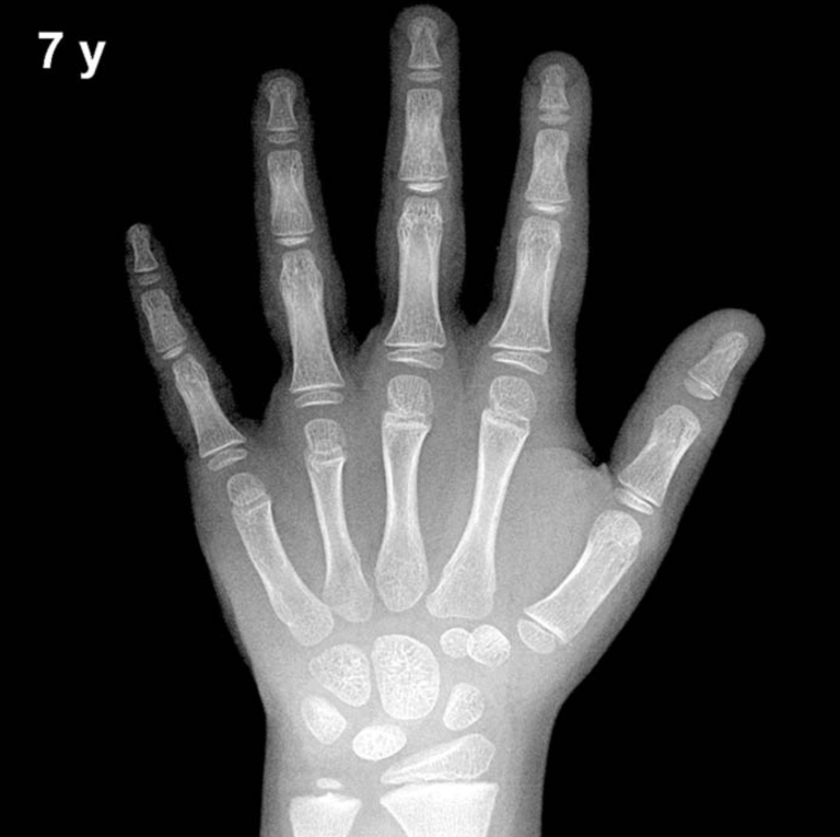
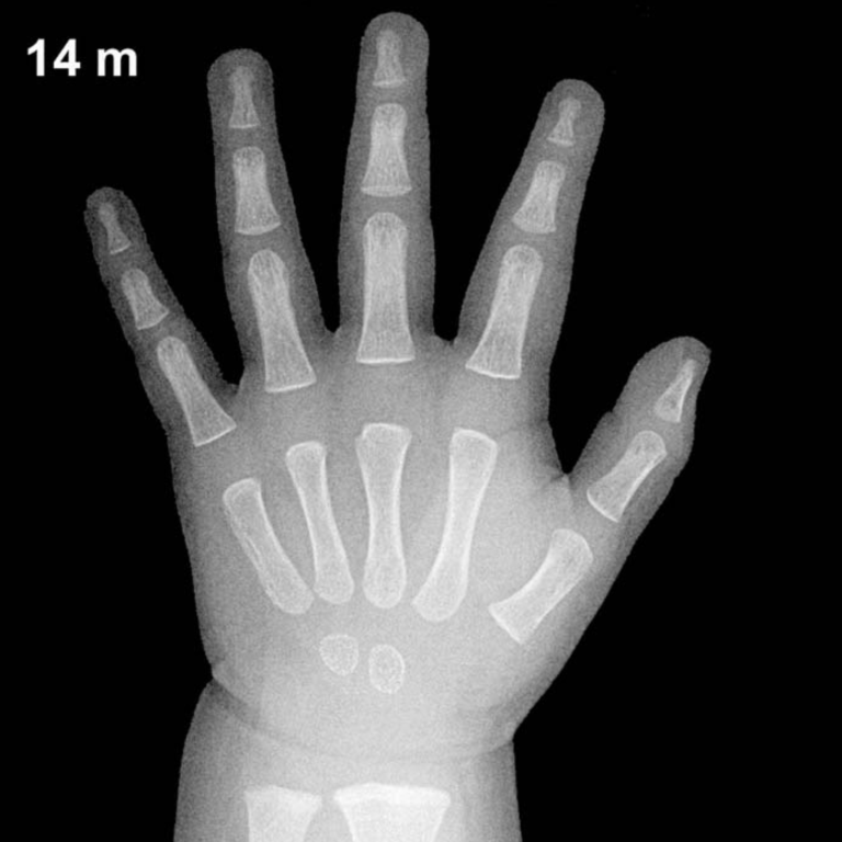
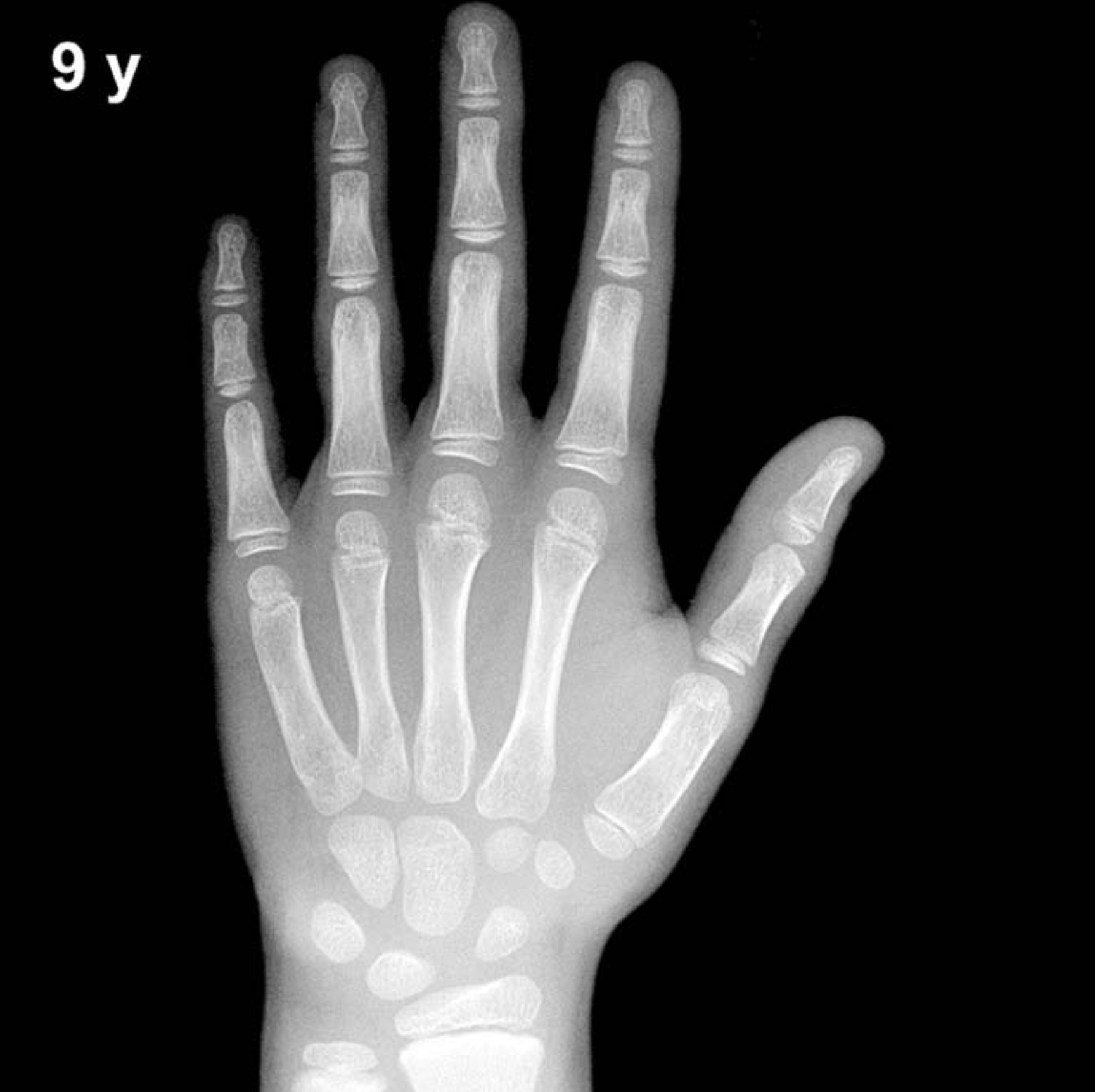
By age 9 in boys, all eight carpal bones are typically ossified. The capitate and hamate appear in early infancy, the triquetral by approximately 2–3 years, the lunate by 3–4 years, the scaphoid, trapezium, and trapezoid between 4–6 years, and the pisiform — which is characteristically late — generally does not appear until approximately 11–14 years in boys, so its absence is entirely normal at this age.
- Epiphyses of all metacarpals and proximal, middle, and distal phalanges are well ossified and show progressive capping and modeling.
- The distal radial epiphysis is well established (typically visible from ~1 year) and by age 9 should show a broad, well-defined plate with early ulnar styloid development.
- The distal ulnar epiphysis is typically present, having appeared between 5–7 years in boys.
- Epiphyseal width relative to metaphyseal diameter and the degree of epiphyseal capping are important maturity indicators at this age.
The overall skeletal pattern at 9 years in boys reflects mid-childhood maturation, prior to the pubertal acceleration. Subtle asymmetry between metacarpal and phalangeal epiphyseal development may help differentiate a bone age near 8 versus 10 years when comparing against adjacent atlas plates.
Clinical Pearls
At age 9, the typical standard deviation for skeletal age in boys is approximately ±1 year; a bone age more than 2 SD outside the mean warrants further evaluation. Girls are skeletally advanced relative to boys by approximately 12–18 months at this chronological age, so normal atlas plates differ meaningfully between sexes. A bone age significantly advanced (e.g., ≥2 years ahead) may suggest central or peripheral precocious puberty, congenital adrenal hyperplasia, or exogenous androgen exposure. Conversely, a bone age significantly delayed raises concern for growth hormone deficiency, hypothyroidism, or constitutional delay of growth and puberty.
A key interpretive pitfall is over-reliance on a single ossification center: skeletal maturity should be assessed globally across multiple bones rather than anchored to any one landmark. Nutritional status, chronic illness, and population differences from the original Greulich-Pyle cohort can all shift expected findings. Reference: Greulich WW, Pyle SI. Radiographic Atlas of Skeletal Development of the Hand and Wrist. 2nd ed. Stanford University Press, 1959.