Bone Age in Girls Aged 6 Years — Greulich-Pyle Hand and Wrist X-Ray Reference
Bone age assessment using a left-hand and wrist radiograph is a cornerstone of pediatric endocrine and growth evaluation. The Greulich-Pyle (GP) method compares a child’s skeletal maturation against standardized atlas plates derived from a North American reference population. In a 6-year-old girl, this assessment is particularly relevant when evaluating conditions such as precocious puberty, growth hormone deficiency, hypothyroidism, or constitutional growth delay.
Expected Ossification Centers and Skeletal Findings
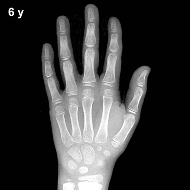
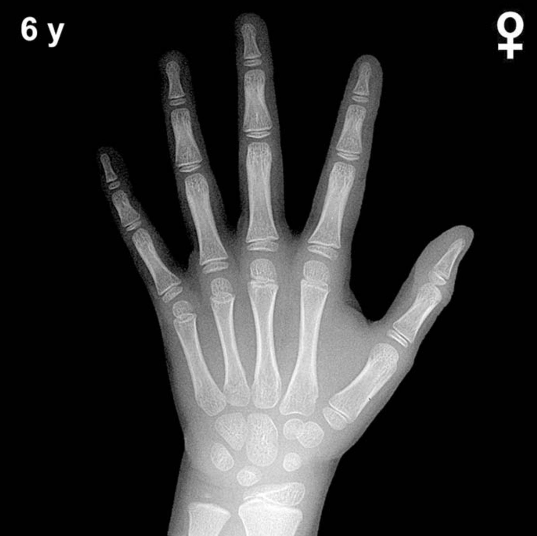
By 6 years of age in girls, all primary carpal ossification centers are typically visible. The capitate (appearing ~3 months) and hamate (~6 months) are well-established and should appear mature in contour. The triquetrum (appearing ~2–3 years), lunate (~3–4 years), and scaphoid, trapezium, and trapezoid (typically ossifying between 4–6 years) should all be present or just completing their initial appearance at this age. The pisiform is not yet expected; in girls it typically appears between 9–12 years.
Epiphyseal development at 6 years in girls includes a well-formed distal radial epiphysis (ossifying from ~1 year), which by this age shows increasing width and definition. The distal ulnar epiphysis typically appears between 5–7 years and may be newly visible or consolidating at this stage. Metacarpal and proximal, middle, and distal phalangeal epiphyses are present and should demonstrate progressive capping of their respective metaphyses. The thumb sesamoid is not expected until the peripubertal period.
- Capitate and hamate: well ossified
- Triquetrum and lunate: established
- Scaphoid, trapezium, trapezoid: present or newly appearing
- Distal radial epiphysis: well formed
- Distal ulnar epiphysis: appearing around this age
- Pisiform and thumb sesamoid: not yet expected
Clinical Pearls
Girls are skeletally advanced compared to boys by approximately 6–12 months at this age, a difference that widens further during puberty. The standard deviation for GP bone age at 6 years is roughly ±1 year, so a bone age between approximately 5 and 7 years is within the normal range for most girls. A bone age advanced by more than 2 SD should prompt evaluation for precocious puberty, congenital adrenal hyperplasia, or exogenous androgen/estrogen exposure. Conversely, a delayed bone age raises concern for growth hormone deficiency, hypothyroidism, or constitutional delay of growth and puberty.
A key interpretive pitfall is population generalizability: the GP atlas was standardized on a mid-20th-century North American cohort of primarily European descent, and skeletal maturation may differ in children of other ethnic backgrounds. Clinical decisions should always integrate bone age findings with auxological data, growth velocity, and biochemical evaluation. Reference: Greulich WW, Pyle SI. Radiographic Atlas of Skeletal Development of the Hand and Wrist. 2nd ed. Stanford University Press, 1959.