Bone Age in Boys Aged 12 Months — Greulich-Pyle Hand and Wrist X-Ray Reference
Bone age assessment using a left hand and wrist radiograph is a well-established method for evaluating skeletal maturity in children, standardized through the Greulich-Pyle (GP) atlas. In infants such as a 12-month-old boy, this assessment helps clinicians detect early deviations in skeletal development that may indicate underlying endocrine, nutritional, or genetic conditions. Accurate bone age estimation at this early stage supports timely intervention in disorders affecting growth.
Expected Ossification Centers and Skeletal Findings
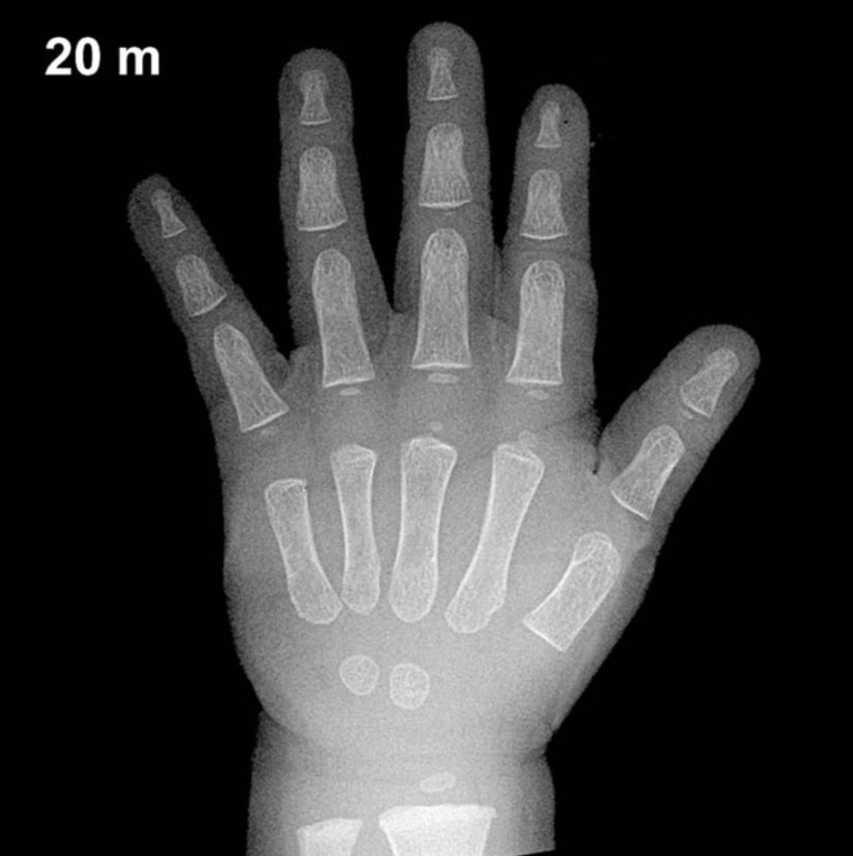
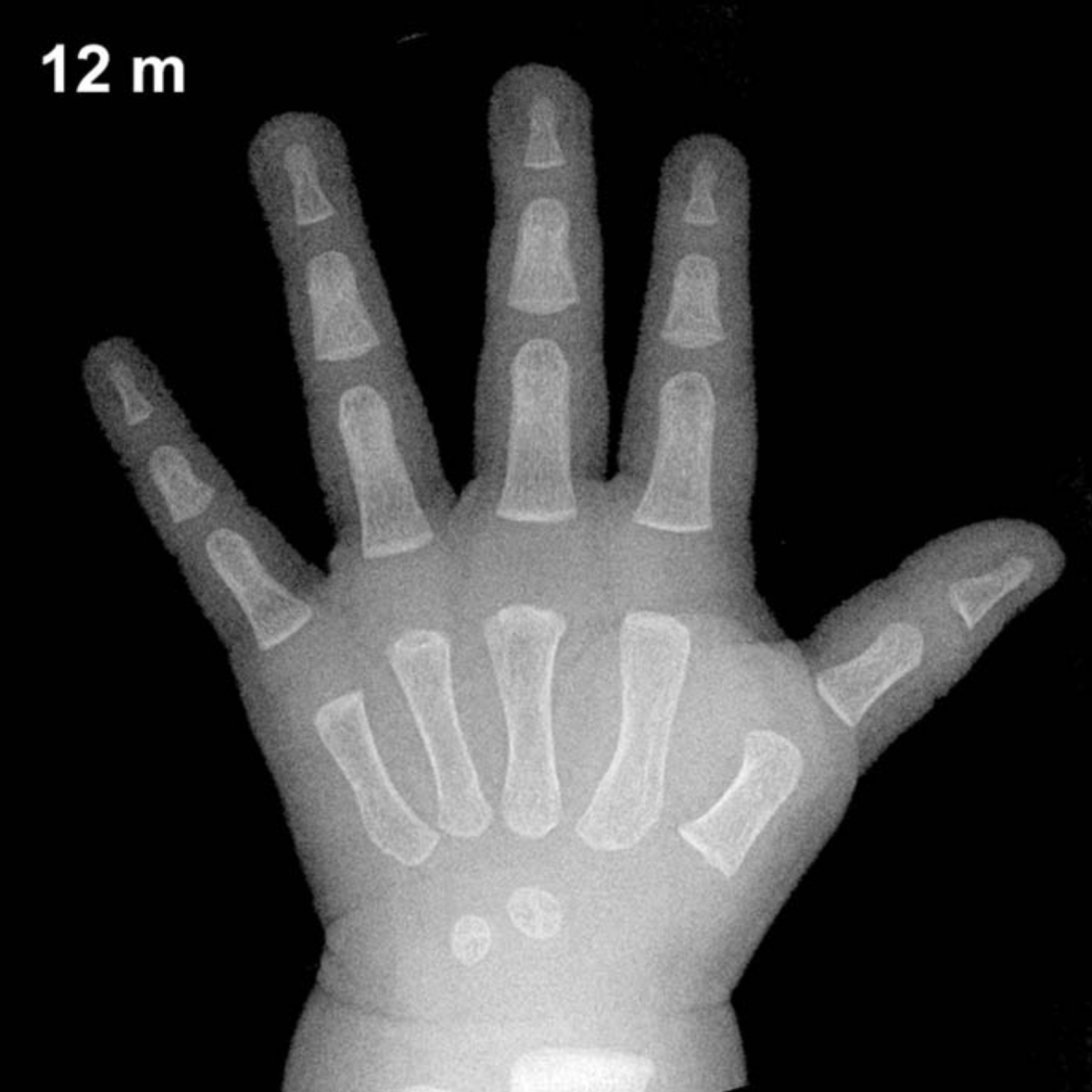
At 12 months of age in boys, skeletal maturation is still in its early stages, and relatively few ossification centers are visible on a hand and wrist radiograph. The capitate is typically the first carpal bone to ossify, appearing as early as 1–3 months of age and should be well established by this age. The hamate generally follows, ossifying around 3–6 months, and is usually visible by 12 months in boys.
By 12 months, the distal radial epiphysis is expected to be present or just beginning to appear, typically ossifying around 9–12 months in boys — making this a key landmark to assess at this age. The remaining carpal bones (triquetral, lunate, scaphoid, trapezium, trapezoid, and pisiform) are not yet expected to be ossified at this stage. The distal ulnar epiphysis and thumb sesamoid are also absent, as these appear considerably later in development.
- Capitate: Present (ossifies ~1–3 months)
- Hamate: Present (ossifies ~3–6 months)
- Distal radial epiphysis: Appearing or just visible (~9–12 months in boys)
- All other carpal centers: Not yet expected
- Distal ulnar epiphysis: Absent (typically appears ~5–7 years)
Clinical Pearls
There is notable physiologic variability in ossification timing even among healthy infants; a range of approximately ±2–3 months is commonly observed at this age. It is important to recognize that girls are skeletally more advanced than boys throughout childhood, with the gap averaging several months during infancy and widening further around puberty. At 12 months, a boy’s bone age that appears significantly advanced — showing more carpal centers than expected — may warrant investigation for conditions such as congenital adrenal hyperplasia or other causes of androgen excess. Conversely, a bone age that is notably delayed, with absent capitate or hamate, may raise concern for hypothyroidism, growth hormone deficiency, or severe nutritional deficiency.
A key interpretive pitfall at this age is over-reliance on a single ossification center: the presence or absence of the distal radial epiphysis alone should be interpreted in the context of overall clinical findings and not used in isolation to assign a bone age. Reference: Greulich WW, Pyle SI. Radiographic Atlas of Skeletal Development of the Hand and Wrist. 2nd ed. Stanford University Press, 1959.