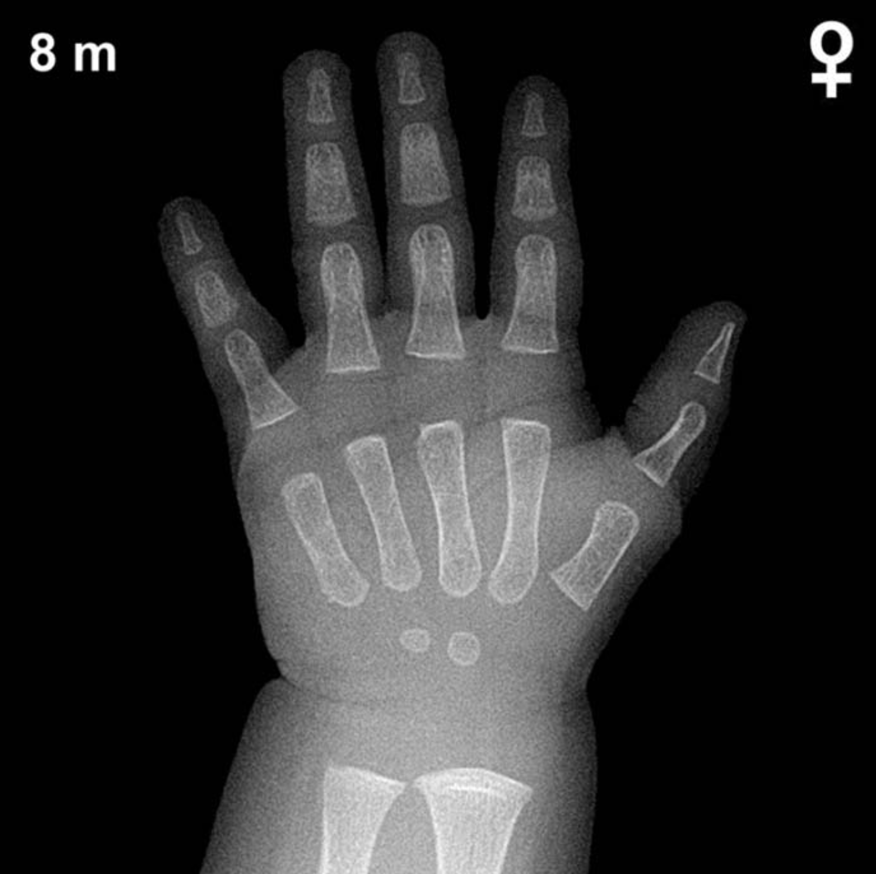
Bone Age in Girls Aged 8 Months — Greulich-Pyle Hand and Wrist X-Ray Reference
Bone age assessment using the Greulich-Pyle (GP) atlas compares a left-hand and wrist radiograph against standardized reference plates to estimate skeletal maturity in children. In infants, this evaluation is particularly valuable in the workup of growth disorders, suspected endocrine abnormalities, and, less commonly, medico-legal age estimation. At 8 months of age in girls, only a small number of ossification centers are expected to be visible, making careful identification of each center critically important.
Expected Ossification Centers and Skeletal Findings
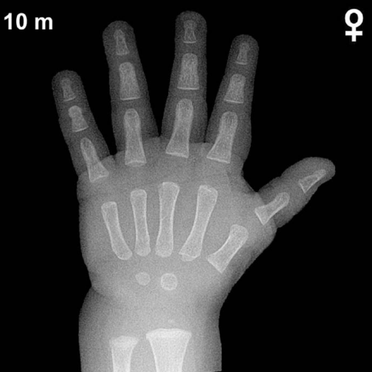
At 8 months of age in girls, the hand and wrist skeleton is at an early stage of ossification. According to the Greulich-Pyle atlas, the capitate is typically the first carpal bone to ossify, appearing at approximately 1–3 months, and should be well established by this age. The hamate follows closely, typically appearing by around 2–6 months, and is also generally visible at 8 months in girls.
Beyond the two medial carpal bones, the remaining carpals are not yet expected to be ossified at this age. The distal radial epiphysis typically begins to appear around 9–12 months in girls, so it may just be emerging or still absent at 8 months. The distal ulnar epiphysis does not ordinarily appear until approximately 5–7 years of age. Epiphyses of the metacarpals and phalanges are generally not yet visible or are only beginning to develop at this stage.
- Capitate: present (typically ossifies ~1–3 months)
- Hamate: present (typically ossifies ~2–6 months)
- Distal radial epiphysis: absent or just appearing (~9–12 months in girls)
- Remaining carpals (triquetral, lunate, scaphoid, trapezium, trapezoid, pisiform): not yet expected
- Metacarpal and phalangeal epiphyses: not yet expected at this age
Clinical Pearls
Skeletal maturation in girls is consistently ahead of boys by approximately 1–2 months during infancy, a gap that widens significantly during puberty. At 8 months, the normal variation in bone age spans roughly ±2–3 months around the chronological age; absence of both the capitate and hamate would suggest notable skeletal delay warranting further evaluation. A bone age significantly advanced beyond 8 months in a female infant may prompt consideration of conditions such as congenital adrenal hyperplasia or other causes of early androgen excess, while marked delay may raise concern for hypothyroidism, growth hormone deficiency, or other metabolic disorders. A key interpretive pitfall at this age is over-reliance on carpal bone size alone; both the presence and morphology of the capitate and hamate should be considered alongside clinical context. Reference: Greulich WW, Pyle SI. Radiographic Atlas of Skeletal Development of the Hand and Wrist. 2nd ed. Stanford University Press, 1959.