Bone Age in Girls Aged 9 Years — Greulich-Pyle Hand and Wrist X-Ray Reference
Bone age assessment using a left-hand and wrist radiograph is a cornerstone of pediatric endocrine and growth evaluation, allowing clinicians to compare skeletal maturation against standardized norms. The Greulich-Pyle method matches the radiograph to the closest atlas standard, providing an estimated skeletal age that may differ from chronological age in pathological or variant states. At 9 years, girls are approaching the early stages of pubertal skeletal maturation, making accurate bone age interpretation particularly relevant for evaluating precocious puberty, adrenal disorders, and growth concerns.
Expected Ossification Centers and Skeletal Findings
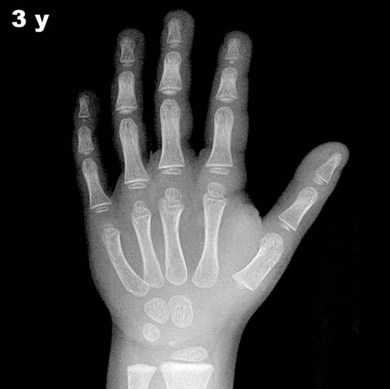
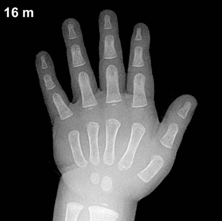
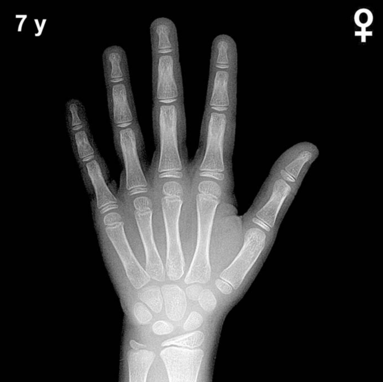
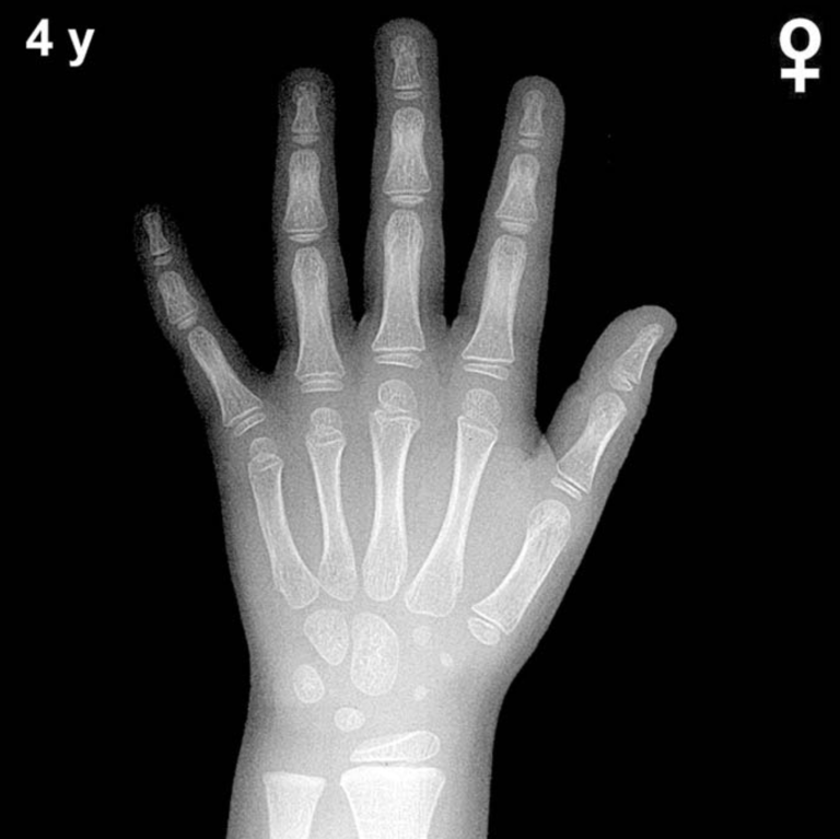
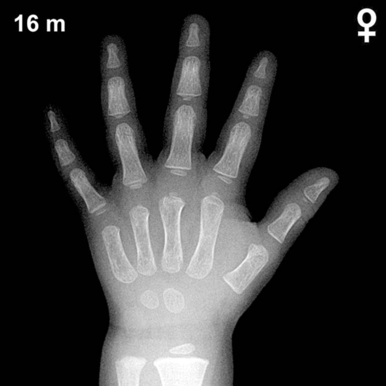
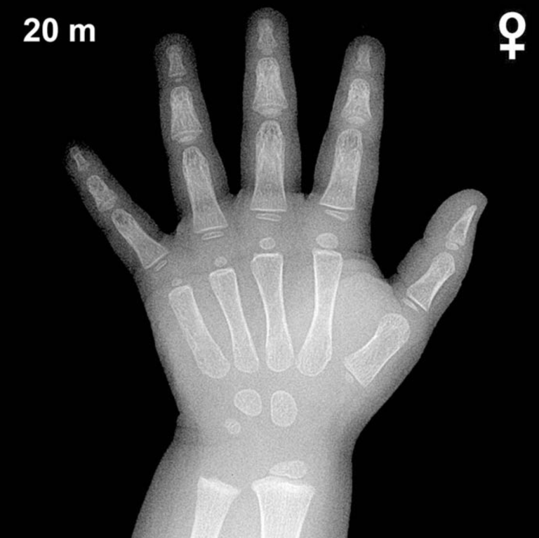
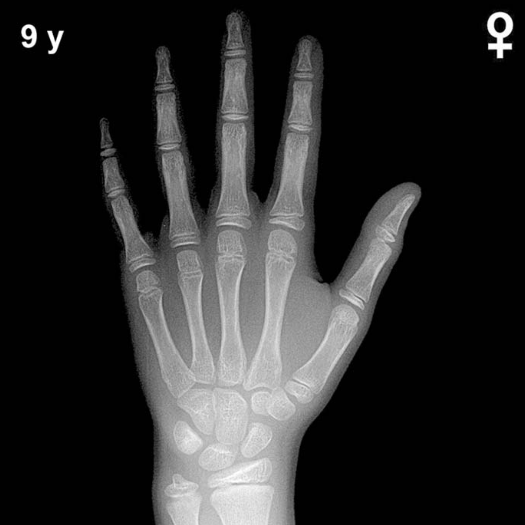
By 9 years of age in girls, all eight carpal bones are typically ossified. The capitate and hamate appear in early infancy, the triquetral by approximately 2–3 years, the lunate by 3–4 years, and the scaphoid, trapezium, and trapezoid between 4–6 years. The pisiform, which ossifies later, is generally expected to be visible in girls around this age, typically appearing between 8–10 years in girls — earlier than in boys.
Epiphyseal maturation is well advanced at 9 years in girls. The distal radial epiphysis is well established, and the distal ulnar epiphysis — which typically appears around 5–7 years — should be clearly visible. Epiphyses of the metacarpals and phalanges show progressive capping and increased density relative to younger ages. The thumb sesamoid (adductor pollicis) is a key peripubertal landmark and may be appearing or just visible at this age in girls, typically emerging around 9–11 years in girls.
- Capitate & hamate: Present since infancy
- Triquetral, lunate: Present since early childhood
- Scaphoid, trapezium, trapezoid: Present since approximately 4–6 years
- Pisiform: Typically visible by 8–10 years in girls
- Distal ulnar epiphysis: Well established
- Thumb sesamoid: May be emerging at this age
Clinical Pearls
Girls’ skeletal maturation consistently runs ahead of boys’ by approximately 1–2 years throughout childhood and adolescence. At 9 years, a bone age advance of more than 2 standard deviations (roughly >2 years ahead) should prompt evaluation for precocious puberty, congenital adrenal hyperplasia, or exogenous androgen/estrogen exposure. Conversely, a bone age more than 2 years delayed may suggest growth hormone deficiency, hypothyroidism, or constitutional delay of growth and puberty. The appearance or absence of the thumb sesamoid can serve as a useful pubertal staging marker at this age.
A key interpretive pitfall is that the Greulich-Pyle atlas was derived from a mid-20th-century North American population of predominantly European descent; secular trends and ethnic variation may influence skeletal maturation, potentially leading to systematic over- or underestimation in diverse populations. Clinical context should always complement radiographic findings. Reference: Greulich WW, Pyle SI. Radiographic Atlas of Skeletal Development of the Hand and Wrist. 2nd ed. Stanford University Press, 1959.