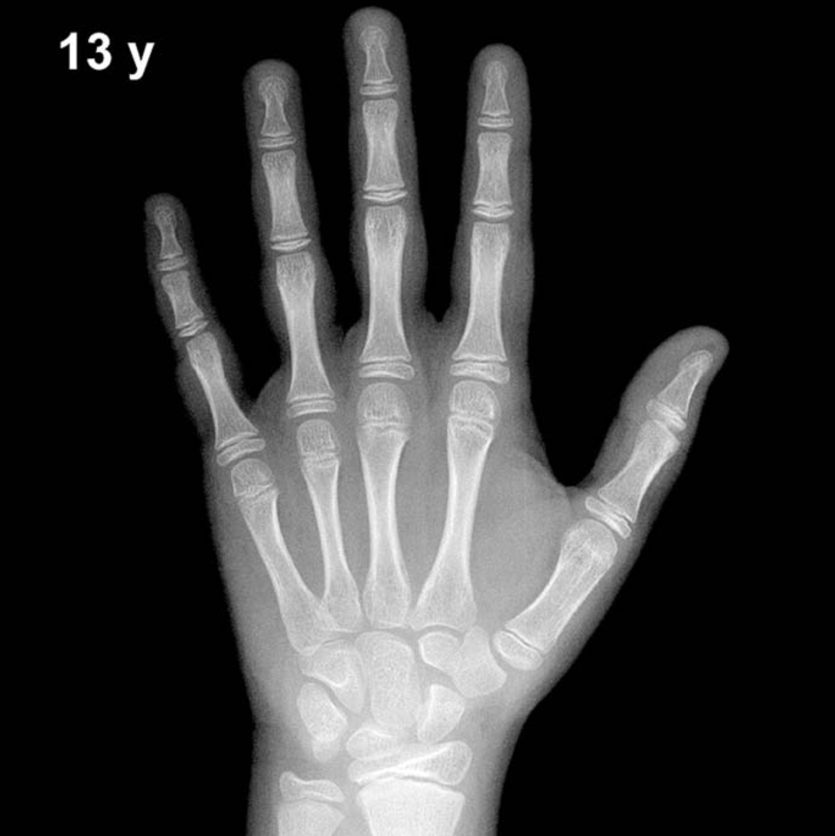
Bone Age in Boys Aged 13 Years — Greulich-Pyle Hand and Wrist X-Ray Reference
Bone age assessment using a left-hand and wrist radiograph is a cornerstone of pediatric endocrine and growth evaluation, allowing clinicians to compare skeletal maturation against established norms. The Greulich-Pyle (GP) atlas provides sex-specific standard radiographs against which a patient’s film is matched to assign a skeletal age. In 13-year-old boys, this assessment is particularly relevant for investigating early or delayed puberty, growth hormone deficiency, and constitutional growth delay.
Expected Ossification Centers and Skeletal Findings
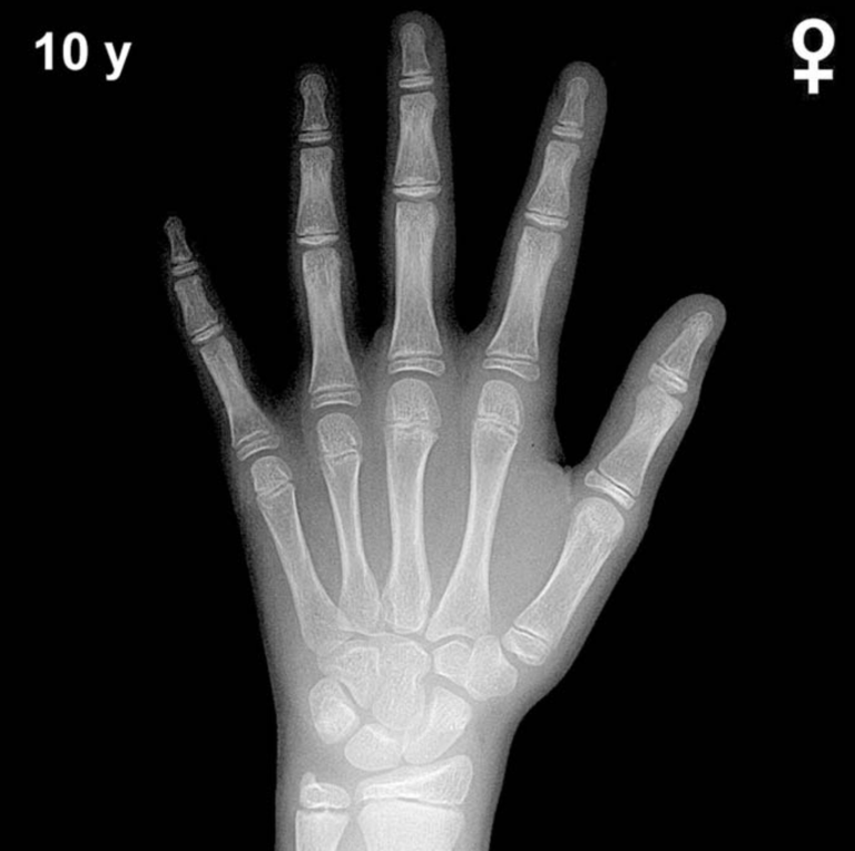
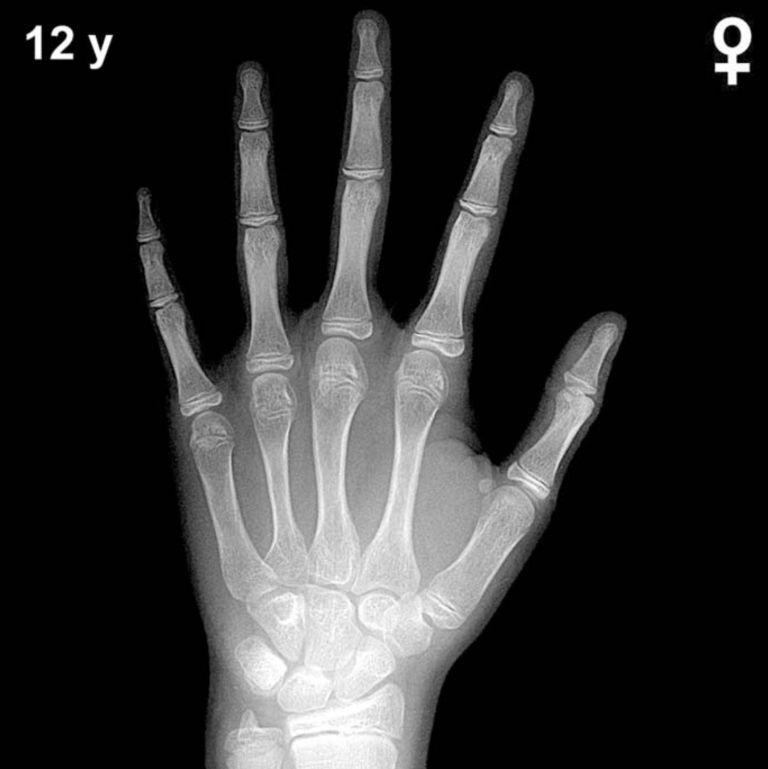
By 13 years of age in boys, all primary carpal ossification centers are well established. The capitate and hamate (appearing at approximately 3 and 6 months, respectively), triquetral, lunate, scaphoid, trapezium, and trapezoid are all fully ossified and maturing in morphology. The pisiform, which typically appears between approximately 11 and 14 years in boys, is expected to be visible or just emerging at this age, making its presence or absence a useful landmark at this stage.
Epiphyseal development is advanced at 13 years. The distal radial and ulnar epiphyses are well formed, with the distal ulnar epiphysis having appeared around age 5–7 years. Metacarpal and phalangeal epiphyses show progressive capping and widening consistent with mid-pubertal maturation. The adductor sesamoid of the thumb typically appears in the peripubertal period (approximately GP bone age 13–14 years in boys), so its presence or absence is a key milestone to assess on films at this chronological age.
- All eight carpal bones: ossified and morphologically maturing
- Pisiform: should be present or emerging by this age in boys
- Thumb adductor sesamoid: appearing around bone age 13–14 years in boys; a critical pubertal marker
- Distal radial and ulnar epiphyses: well developed, progressive fusion not yet expected
- Metacarpal/phalangeal epiphyses: broad, cap-like morphology consistent with active pubertal growth
Clinical Pearls
Girls’ skeletal maturation is typically 1–2 years ahead of boys at comparable chronological ages, so GP standards are strictly sex-specific. At 13 years in boys, a bone age more than 2 standard deviations advanced (≥15 years) may prompt evaluation for precocious puberty or androgen excess, while a bone age significantly delayed (≤11 years) raises concern for growth hormone deficiency, hypothyroidism, or constitutional delay of growth and puberty. In phenotypic females with a 46,XY karyotype, using male standards is essential. A key interpretive pitfall is over-reliance on a single bone match: GP assessment has an inter-observer variability of approximately ±6–12 months, and clinical context must always guide interpretation. Reference: Greulich WW, Pyle SI. Radiographic Atlas of Skeletal Development of the Hand and Wrist. 2nd ed. Stanford University Press, 1959.