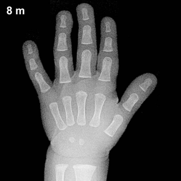
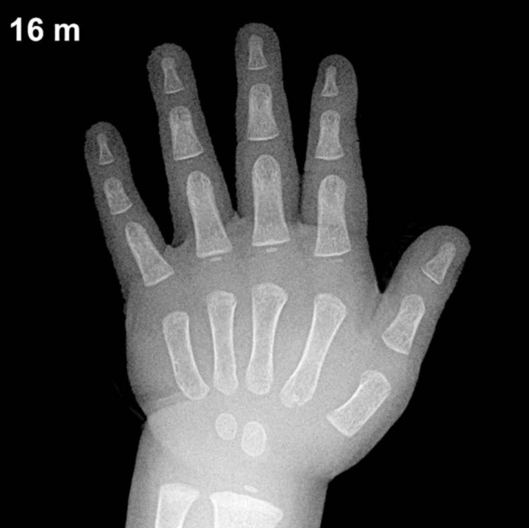
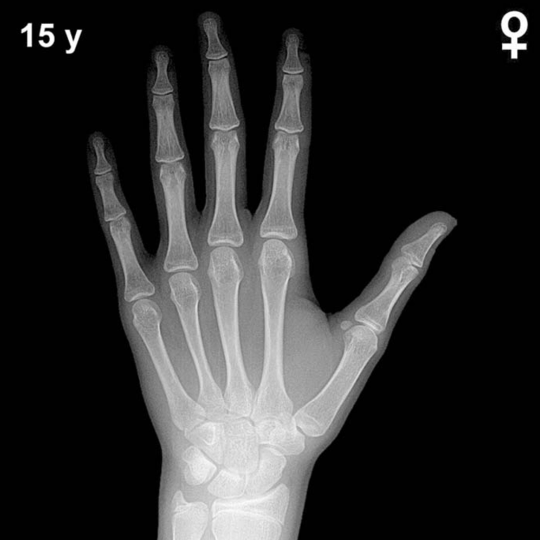
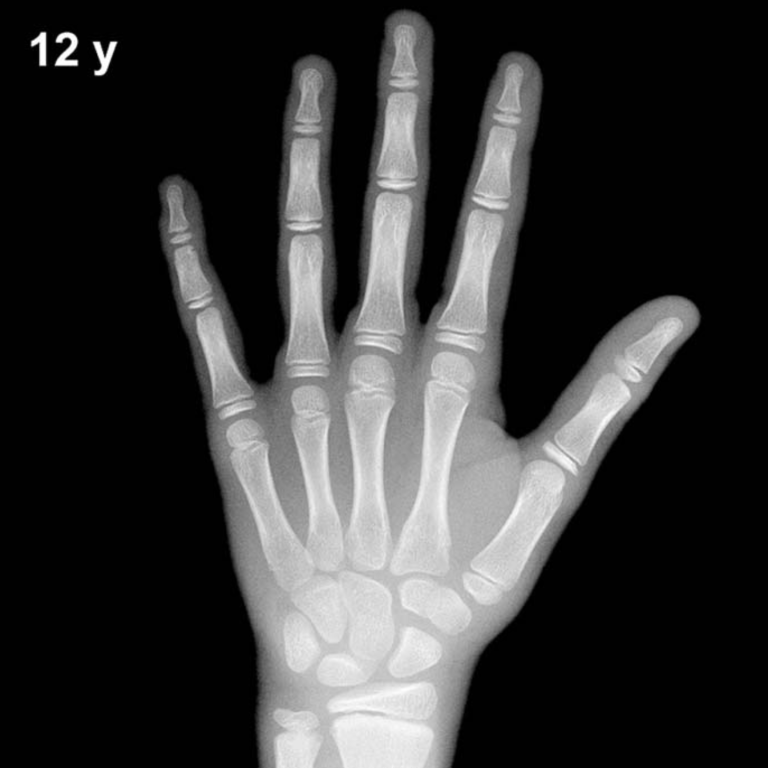
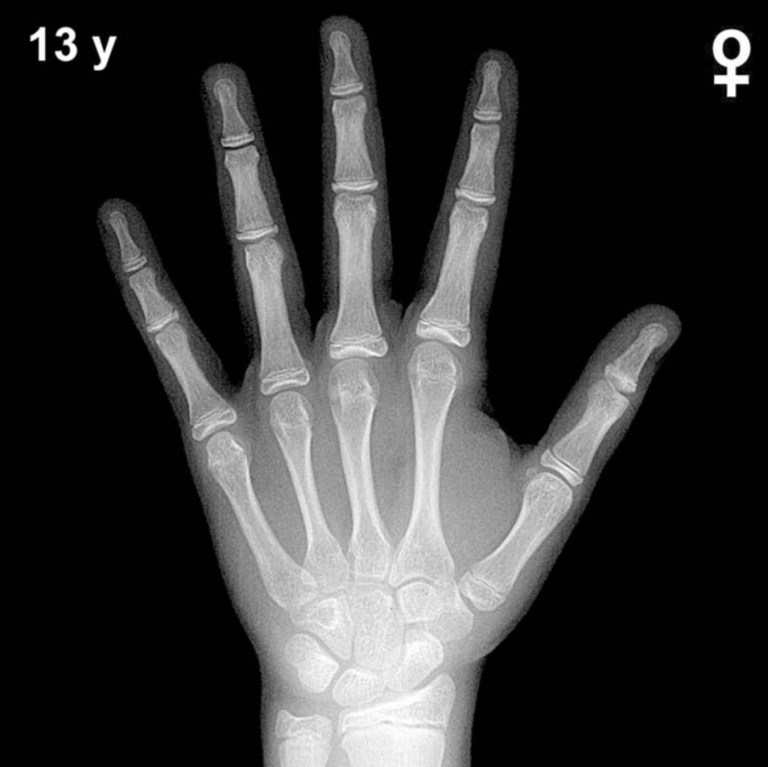
Bone Age in Boys Aged 16 Years — Greulich-Pyle Hand and Wrist X-Ray Reference
Bone age assessment using a left-hand and wrist radiograph is a cornerstone of pediatric endocrine and growth evaluation, comparing a child’s skeletal maturity against established norms. The Greulich-Pyle atlas remains the most widely used reference standard, providing sex-specific atlas plates derived from longitudinal radiographic data. In 16-year-old boys, bone age evaluation is particularly relevant in the workup of delayed or precocious puberty, constitutional growth delay, and hypogonadism.
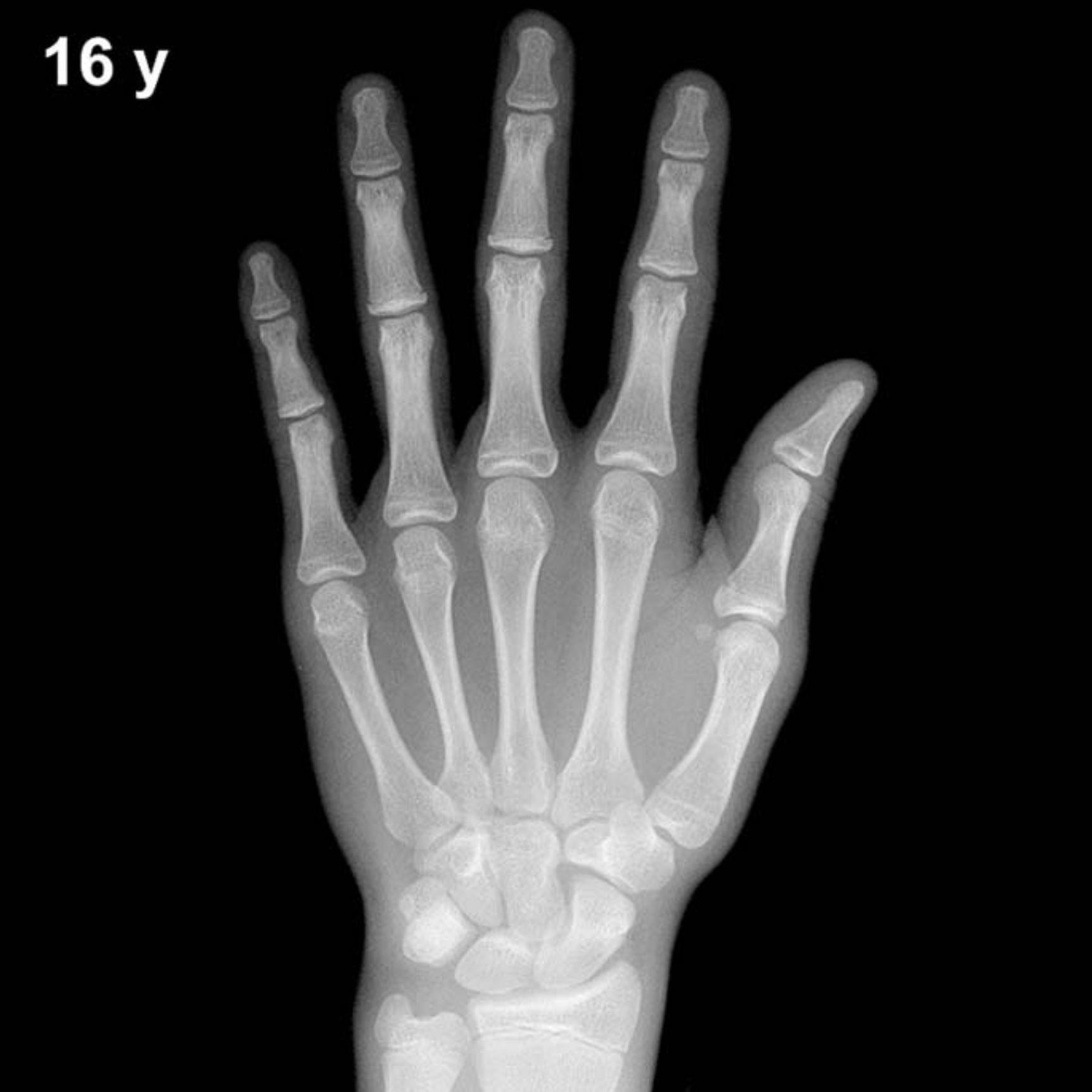
Expected Ossification Centers and Skeletal Findings
By 16 years in boys, virtually all ossification centers of the hand and wrist are well established. All eight carpal bones — capitate, hamate, triquetral, lunate, scaphoid, trapezium, trapezoid, and pisiform — should be fully ossified. The pisiform, which typically appears between approximately 11 and 14 years in boys, is expected to be clearly visible by this age.
Epiphyseal development is advanced at 16 years. The distal radial and ulnar epiphyses are well formed and broad. A key milestone at this age is the progressive fusion of epiphyses to their metaphyses: epiphyseal fusion at the proximal and middle phalanges and metacarpals is typically underway or nearing completion, though full fusion of the distal radius may not yet be complete in all boys at exactly 16 years. The adductor sesamoid of the thumb, a reliable pubertal marker, should be present and well ossified by this age.
- All carpal bones: ossified and well corticated
- Pisiform: present and fully ossified
- Thumb sesamoid: present (peripubertal landmark, typically appears ~13–14 yr in boys)
- Phalangeal and metacarpal epiphyses: fusing or nearly fused
- Distal radial epiphysis: broad; fusion in progress but may not be complete
Clinical Pearls
At 16 years, girls’ skeletal maturity is typically 12–18 months ahead of boys of the same chronological age, reflecting the well-documented sex difference in skeletal maturation. A bone age significantly delayed beyond 16 years in boys (e.g., ≥2 years behind) raises consideration of constitutional delay of growth and puberty, growth hormone deficiency, hypothyroidism, or hypogonadism. Conversely, a bone age substantially advanced for chronological age may suggest prior androgen excess, such as congenital adrenal hyperplasia. Standard deviation for bone age assessment at this age is approximately ±1 year, so modest discrepancies require clinical correlation rather than isolated interpretation. A key pitfall is over-reliance on a single atlas plate: the Greulich-Pyle method performs best when the overall pattern of maturation — not one isolated center — guides the assessment. Reference: Greulich WW, Pyle SI. Radiographic Atlas of Skeletal Development of the Hand and Wrist. 2nd ed. Stanford University Press, 1959.