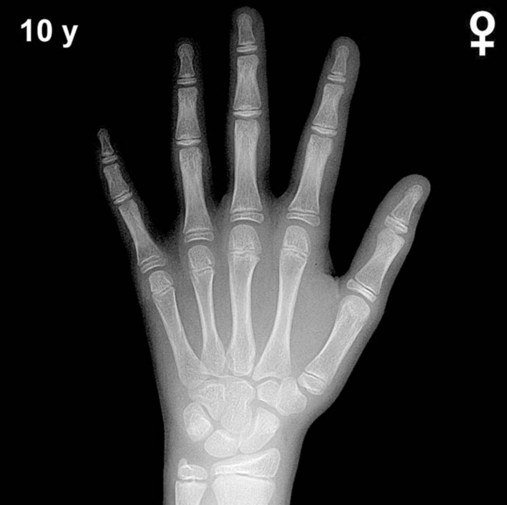
Bone Age in Girls Aged 10 Years — Greulich-Pyle Hand and Wrist X-Ray Reference
Bone age assessment using a left-hand and wrist radiograph is a cornerstone of pediatric endocrine and growth evaluation, comparing a child’s skeletal maturity against standardized atlas references. The Greulich-Pyle method matches the radiograph to the closest atlas standard to estimate skeletal age, which may differ meaningfully from chronological age. This assessment is clinically essential when evaluating precocious or delayed puberty, growth hormone deficiency, hypothyroidism, and other conditions affecting skeletal maturation.
Expected Ossification Centers and Skeletal Findings
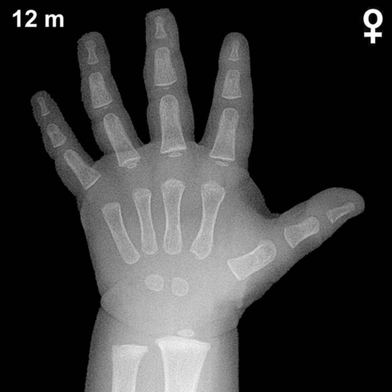
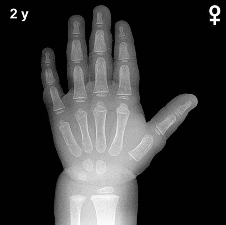
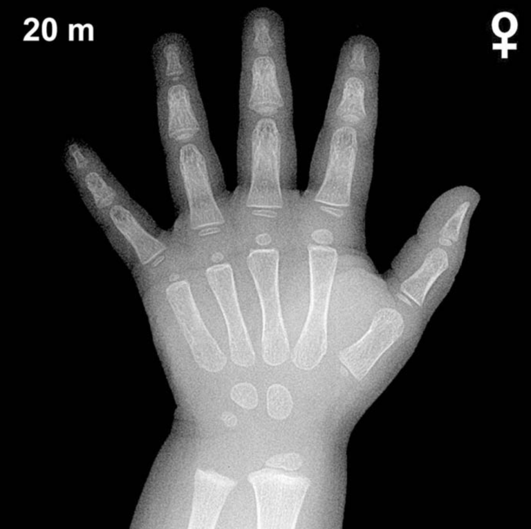
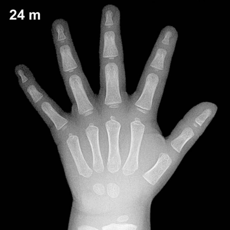
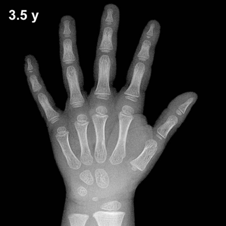
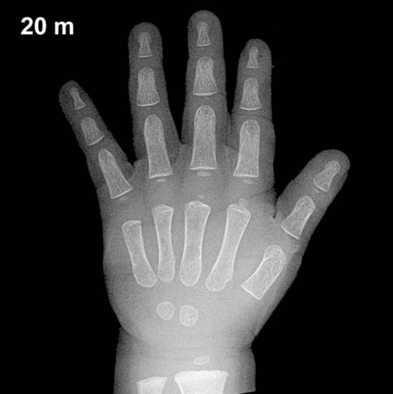
By 10 years of age in girls, all eight carpal bones are typically ossified. The capitate and hamate appear in early infancy, the triquetrum by approximately 2–3 years, the lunate by 3–4 years, the scaphoid, trapezium, and trapezoid between 4–6 years, and the pisiform typically between 9–12 years in girls — meaning the pisiform ossification center is often visible or just appearing at this age, making it an important landmark to assess.
The distal radial epiphysis, present since approximately 1 year of age, is now well-developed and broader. The distal ulnar epiphysis, typically appearing between 5–7 years, should be clearly visible. Epiphyses of the metacarpals and phalanges are well-formed, and fusion has not yet begun. At 10 years, girls are typically in mid-to-late prepubertal or early pubertal skeletal development; the adductor sesamoid of the thumb may be beginning to appear around this age, as its ossification is closely tied to the onset of puberty and is often seen in girls between 10–12 years.
- All eight carpal centers present; pisiform ossification expected
- Distal radial and ulnar epiphyses well established
- Metacarpal and phalangeal epiphyses prominent, unfused
- Adductor sesamoid of thumb may be emerging — a pubertal marker
Clinical Pearls
At 10 years, girls’ skeletal maturity is typically approximately 1–2 years ahead of boys of the same chronological age, reflecting the well-established sex difference in skeletal maturation. A bone age significantly advanced beyond 10 years in a girl of this age raises concern for precocious puberty or exogenous androgen/estrogen exposure, while a notably delayed bone age may suggest growth hormone deficiency, hypothyroidism, or constitutional delay of growth and puberty. Turner syndrome (45,X) may also present with modestly delayed bone age alongside short stature.
A key interpretive pitfall is over-relying on a single skeletal indicator; bone age should be assessed as a composite of multiple ossification and epiphyseal features rather than any one center alone. Population and ethnicity-based variation also exists, and the Greulich-Pyle atlas was standardized on a mid-twentieth century North American cohort. Reference: Greulich WW, Pyle SI. Radiographic Atlas of Skeletal Development of the Hand and Wrist. 2nd ed. Stanford University Press, 1959.