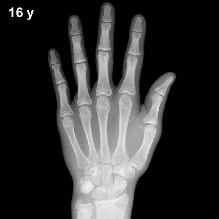
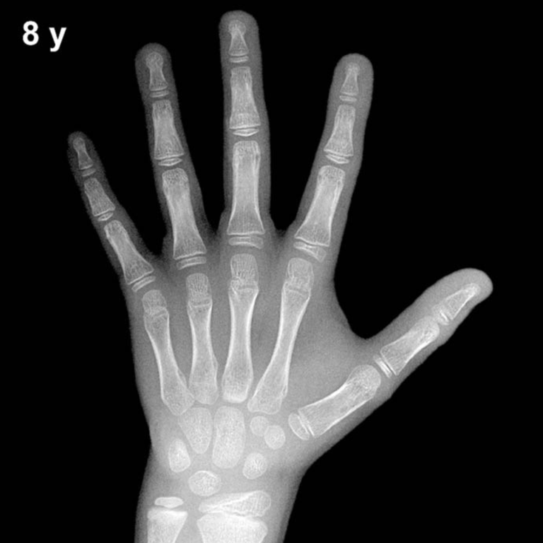
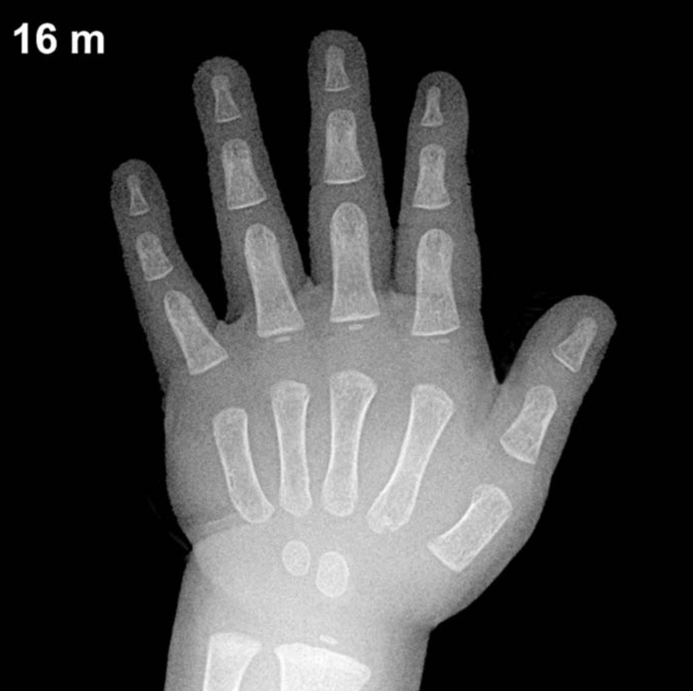
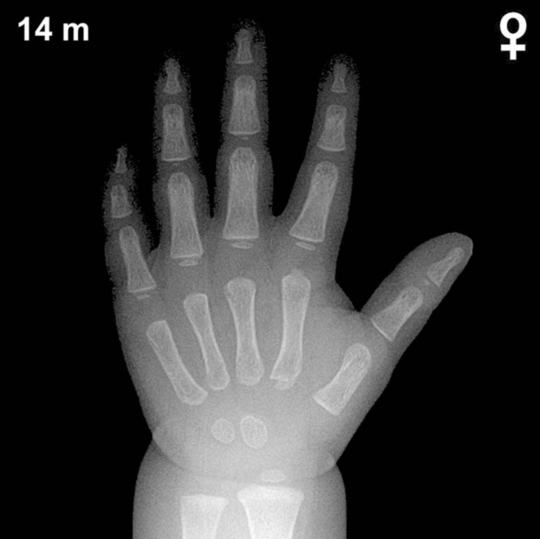
Bone Age in Boys Aged 3.5 Years — Greulich-Pyle Hand and Wrist X-Ray Reference
Bone age assessment using the Greulich-Pyle (GP) atlas compares the skeletal maturation of a child’s left hand and wrist radiograph against standardized reference plates derived from a mid-twentieth-century North American population. In boys aged 3.5 years, this evaluation helps clinicians identify deviations from expected skeletal development that may signal underlying endocrine, metabolic, or constitutional conditions. Accurate bone age estimation is essential in the workup of growth disorders, precocious or delayed puberty, and, in some contexts, forensic age estimation.
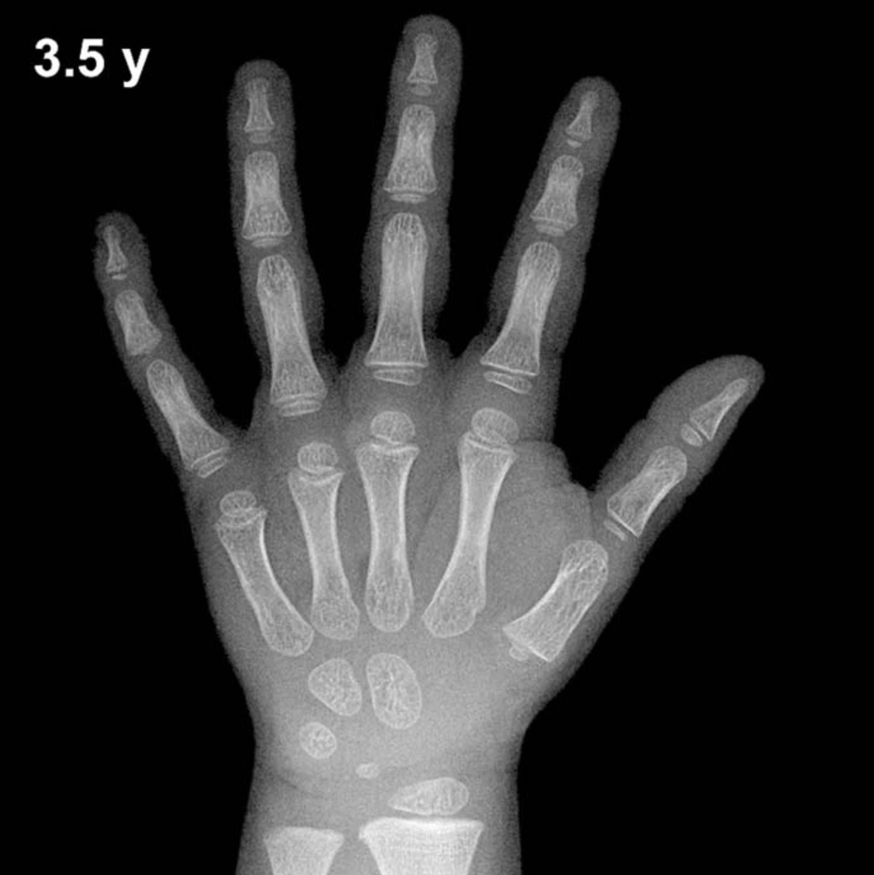
Expected Ossification Centers and Skeletal Findings
By 3.5 years of age in boys, several carpal ossification centers should be visible on a properly exposed hand and wrist radiograph. The capitate and hamate are the earliest carpal bones to ossify, typically appearing within the first year of life (capitate ~3 months, hamate ~6 months), and are well established by this age. The triquetrum typically ossifies between 2 and 3 years in boys and should be present or just appearing at 3.5 years. The lunate ossification center typically emerges between 3 and 4 years, meaning it may be present but is not always fully established at this age.
The distal radial epiphysis is normally well ossified by approximately 1 year and appears as a well-defined, smoothly marginated ossification center at this age. The scaphoid, trapezium, and trapezoid typically ossify between 4 and 6 years in boys and are generally not yet expected at 3.5 years. The distal ulnar epiphysis typically appears between 5 and 7 years and is usually absent at this age. The pisiform and the thumb sesamoid are both considerably later milestones, not expected until late childhood or peripubertal years.
- Present and expected: Capitate, hamate, triquetrum, lunate (emerging), distal radial epiphysis
- Not yet expected: Scaphoid, trapezium, trapezoid, pisiform, distal ulnar epiphysis, thumb sesamoid
Clinical Pearls
At 3.5 years, boys’ skeletal maturity typically lags behind girls of the same chronological age by approximately 6–12 months, a well-documented sex difference that widens further during puberty. A bone age more than 2 standard deviations (roughly ±1.5–2 years at this age) above or below the chronological age warrants clinical attention. Advanced bone age in a 3.5-year-old boy may suggest precocious puberty, congenital adrenal hyperplasia, or exogenous androgen exposure. Delayed bone age raises consideration of growth hormone deficiency, hypothyroidism, or constitutional delay of growth and puberty. A key interpretive pitfall is over-reliance on a single ossification center: normal biological variability means that the overall pattern of maturation across multiple centers should guide assessment rather than any one landmark in isolation.
Reference: Greulich WW, Pyle SI. Radiographic Atlas of Skeletal Development of the Hand and Wrist. 2nd ed. Stanford University Press, 1959.