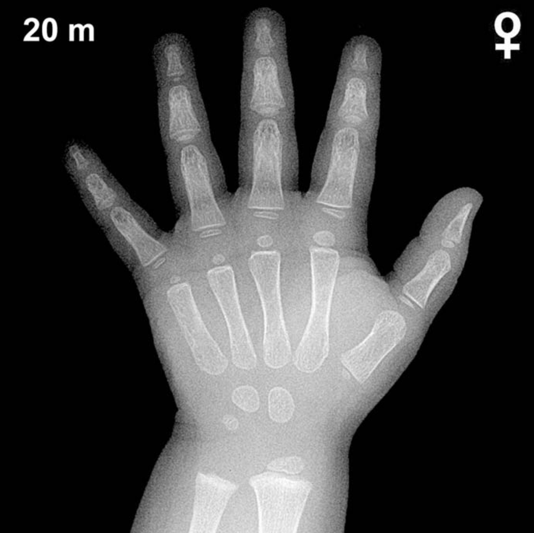
Bone Age in Girls Aged 20 Months — Greulich-Pyle Hand and Wrist X-Ray Reference
Bone age assessment using a posteroanterior radiograph of the left hand and wrist is a standard tool for evaluating skeletal maturity in children. The Greulich-Pyle method compares the child’s radiograph against standardized atlas plates to determine whether skeletal development is appropriate for chronological age. In girls aged 20 months, this assessment is particularly relevant when investigating growth faltering, endocrine disorders such as hypothyroidism, or precocious puberty.
Expected Ossification Centers and Skeletal Findings
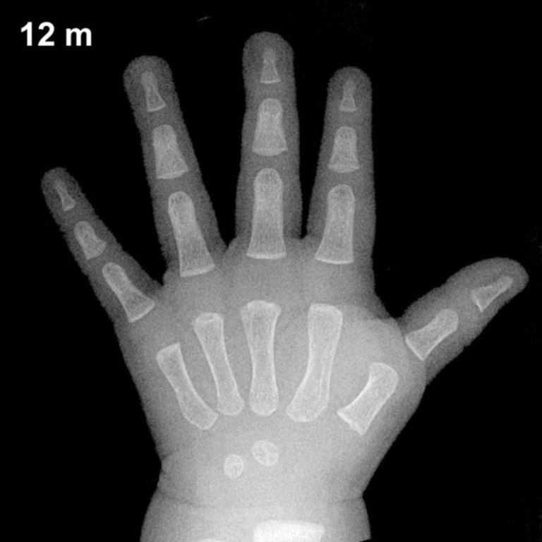
By 20 months of age in girls, several carpal and epiphyseal ossification centers are typically present. The capitate and hamate are among the earliest carpal bones to ossify, appearing around 3 and 6 months of age respectively, and should be well established by this age. The triquetral ossification center typically appears between 2 and 3 years in girls, so it may be absent or just emerging at 20 months.
The distal radial epiphysis typically ossifies around 12 months and should be visible and reasonably well defined by 20 months in girls. The lunate, scaphoid, trapezium, and trapezoid are not expected to be ossified at this age. The distal ulnar epiphysis, pisiform, and thumb sesamoid are also absent at this stage, as these appear considerably later in development.
- Present by 20 months (girls): Capitate, hamate, distal radial epiphysis
- Possibly emerging: Triquetral (may appear around 2–3 years)
- Not yet expected: Lunate, scaphoid, trapezium, trapezoid, pisiform, distal ulnar epiphysis, thumb sesamoid
Clinical Pearls
Skeletal maturation in girls is known to run approximately 2–6 months ahead of boys at this early age, a difference that widens further through mid-childhood. At 20 months, a bone age significantly advanced beyond chronological age may raise concern for precocious puberty or exogenous androgen exposure, while a markedly delayed bone age warrants evaluation for hypothyroidism, growth hormone deficiency, or nutritional deficiency. A bone age delay of 2 or more standard deviations from the mean is generally considered clinically significant. One important pitfall is the limited number of atlas reference plates available for very young children in the Greulich-Pyle atlas, which can reduce precision of assessment in infants and toddlers; clinical correlation and serial imaging are strongly recommended in this age group.
Reference: Greulich WW, Pyle SI. Radiographic Atlas of Skeletal Development of the Hand and Wrist. 2nd ed. Stanford University Press, 1959.