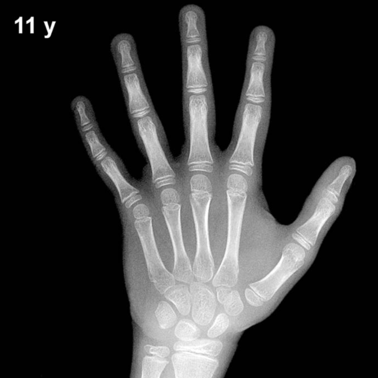
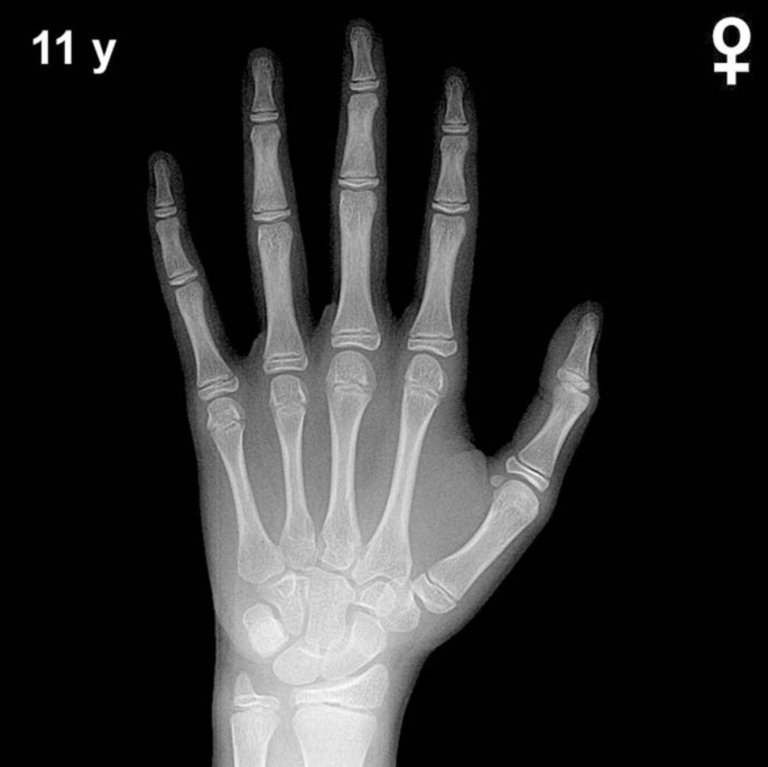
Bone Age in Girls Aged 16 Months — Greulich-Pyle Hand and Wrist X-Ray Reference
Bone age assessment using a left hand and wrist radiograph is a fundamental tool for evaluating skeletal maturity in children. The Greulich-Pyle method compares a child’s radiograph against standard atlas plates to determine whether skeletal development is concordant with chronological age. In girls aged 16 months, this assessment is particularly relevant when investigating growth disorders, thyroid dysfunction, or early signs of endocrine pathology.
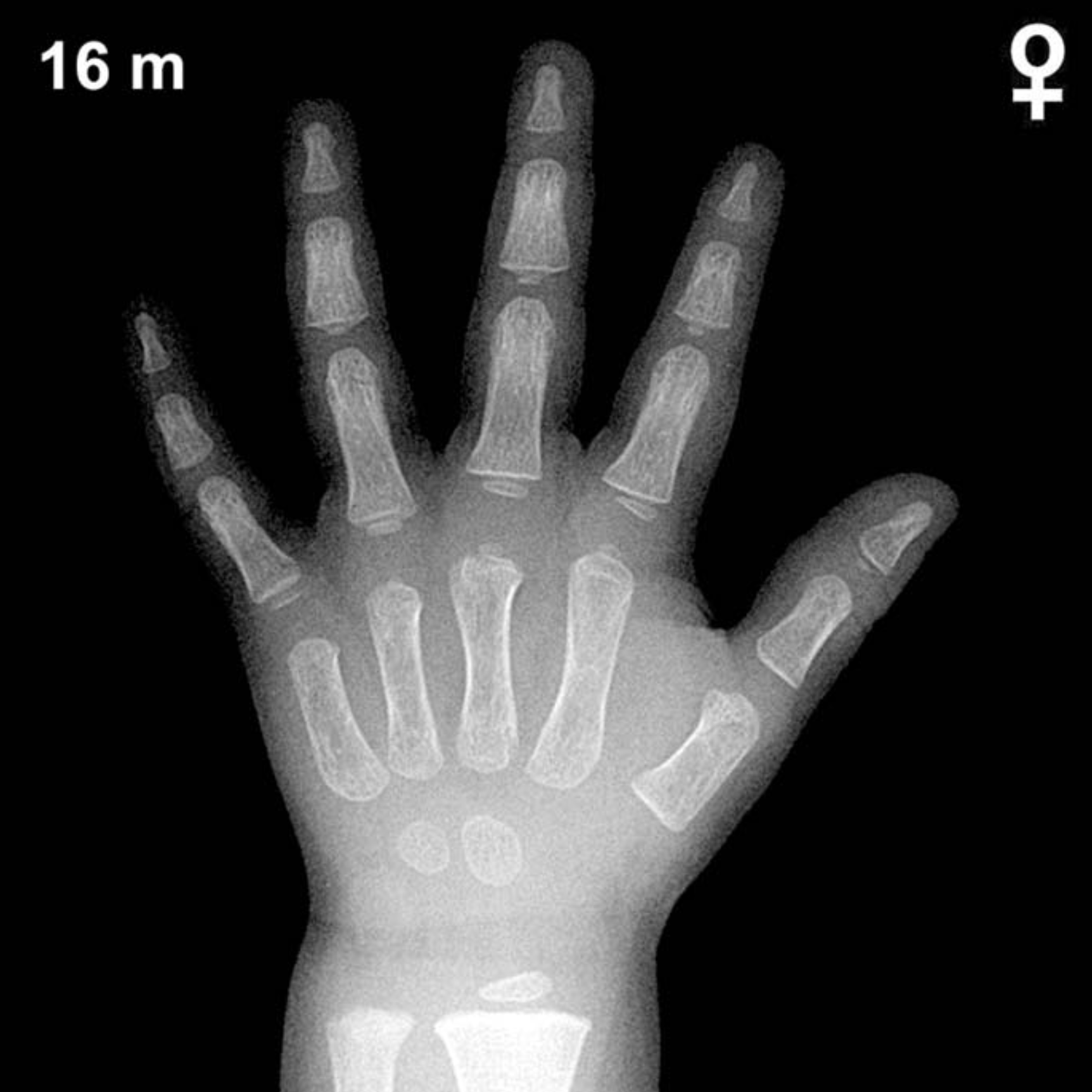
Expected Ossification Centers and Skeletal Findings
By 16 months of age in girls, the earliest carpal ossification centers are typically already present. The capitate and hamate are the first carpals to ossify, appearing at approximately 3 months and 6 months of age respectively, and should be well established by this age. The distal radial epiphysis typically appears around 12 months and is generally visible by 16 months in girls, though its size and definition may still be relatively small.
At this age, most remaining carpal bones — including the triquetral, lunate, scaphoid, trapezium, and trapezoid — are not yet expected to be ossified. The triquetral typically appears between 2 and 3 years in girls, while the lunate follows around 3 to 4 years. The pisiform, sesamoids, and distal ulnar epiphysis are all well beyond the expected window for this age group. Epiphyseal ossification centers of the metacarpals and phalanges may be beginning to emerge at this stage, though development varies by individual bone.
- Present: Capitate, hamate, distal radial epiphysis (emerging)
- Not yet expected: Triquetral, lunate, scaphoid, trapezium, trapezoid, pisiform, distal ulnar epiphysis, thumb sesamoid
Clinical Pearls
Girls demonstrate skeletal advancement relative to boys throughout childhood, typically by several months at this early age. Considerable normal variability exists, and a bone age within approximately ±2 standard deviations of chronological age is generally considered within the normal range. A bone age significantly advanced beyond 16 months in a girl of this chronological age may raise concern for precocious puberty or excess androgen exposure, while a notably delayed bone age may suggest hypothyroidism, growth hormone deficiency, or constitutional delay of growth and development. A key interpretive pitfall at this young age is the limited number of ossification centers available for comparison, which can reduce precision; minor discrepancies should always be interpreted alongside clinical and auxological data rather than in isolation. Reference: Greulich WW, Pyle SI. Radiographic Atlas of Skeletal Development of the Hand and Wrist. 2nd ed. Stanford University Press, 1959.