Bone Age in Boys Aged 8 Months — Greulich-Pyle Hand and Wrist X-Ray Reference
Bone age assessment using a left-hand and wrist radiograph is a standard tool for evaluating skeletal maturity in children, with the Greulich-Pyle atlas serving as the most widely used reference in clinical practice. In infants, this assessment is particularly valuable when investigating endocrine or metabolic conditions that may accelerate or delay normal skeletal development. Even at 8 months of age, the pattern and number of visible ossification centers can offer meaningful clinical information.
Expected Ossification Centers and Skeletal Findings
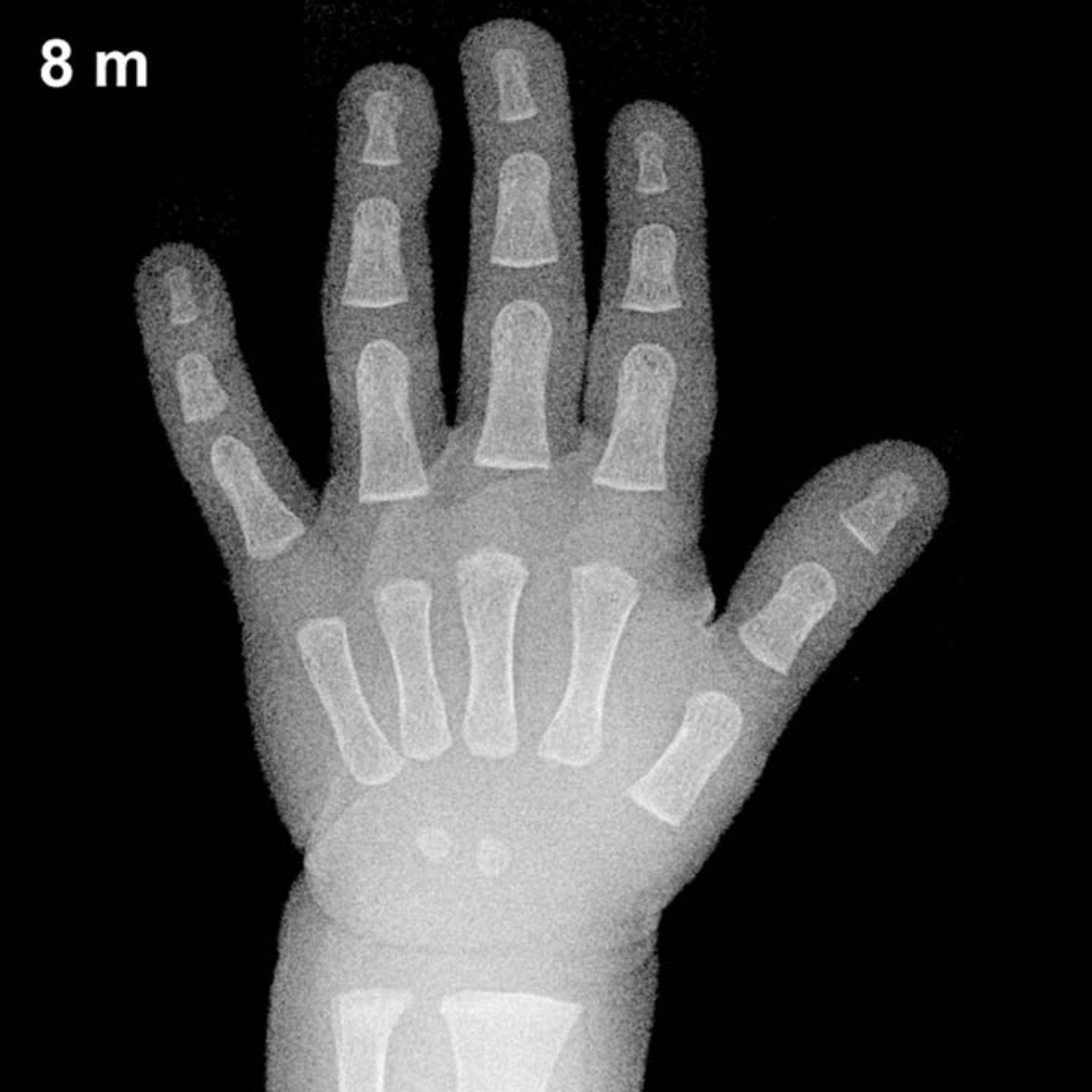
At 8 months of age in boys, the Greulich-Pyle atlas anticipates a limited but characteristic set of ossification centers in the hand and wrist. The capitate is typically the first carpal bone to ossify, appearing around 1–3 months in both sexes, and should be well established by this age. The hamate typically follows closely, ossifying by approximately 3–6 months, and is generally visible at 8 months in boys, though it may appear slightly less mature than in girls of the same age.
Beyond the carpals, the distal radial epiphysis is a key landmark expected to appear around 9–12 months in boys, meaning it may just be emerging or not yet visible at 8 months. The metacarpal and proximal phalangeal epiphyses are generally not yet ossified at this age. No other carpal centers — including the triquetrum, lunate, scaphoid, trapezium, trapezoid, or pisiform — are expected to be present in an 8-month-old boy.
- Capitate: present, well-formed
- Hamate: typically present by 8 months
- Distal radial epiphysis: may be absent or just emerging
- All other carpal centers: not yet expected
Clinical Pearls
Normal skeletal maturation in infants carries significant inter-individual variability; a difference of 1–2 months from the atlas standard is generally considered within acceptable limits at this age. Girls are skeletally more advanced than boys throughout childhood, a difference that becomes more pronounced with age but is already present in infancy. At 8 months, the presence of more than the expected carpal centers — for example, early appearance of the triquetrum — may raise concern for conditions causing accelerated bone age such as congenital adrenal hyperplasia or other causes of excess androgen exposure. Conversely, absence of the capitate and hamate beyond expected age windows may suggest hypothyroidism, hypopituitarism, or other causes of delayed skeletal maturation warranting further workup.
A common pitfall in infant bone age assessment is the reliance on center count alone without considering center size, shape, and density, all of which contribute to the overall maturity rating in the Greulich-Pyle method. Reference: Greulich WW, Pyle SI. Radiographic Atlas of Skeletal Development of the Hand and Wrist. 2nd ed. Stanford University Press, 1959.