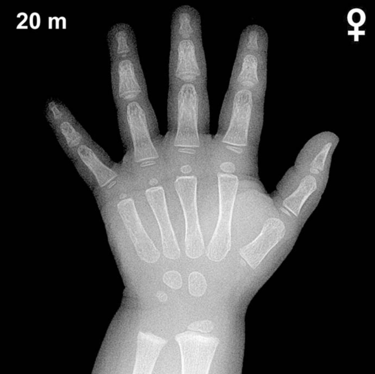
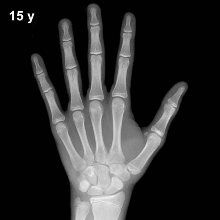
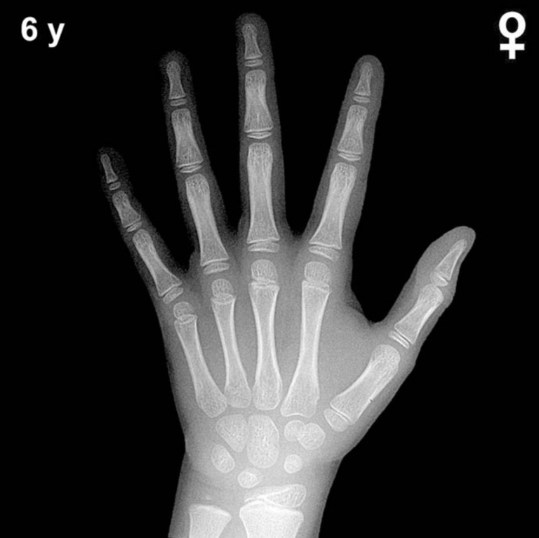
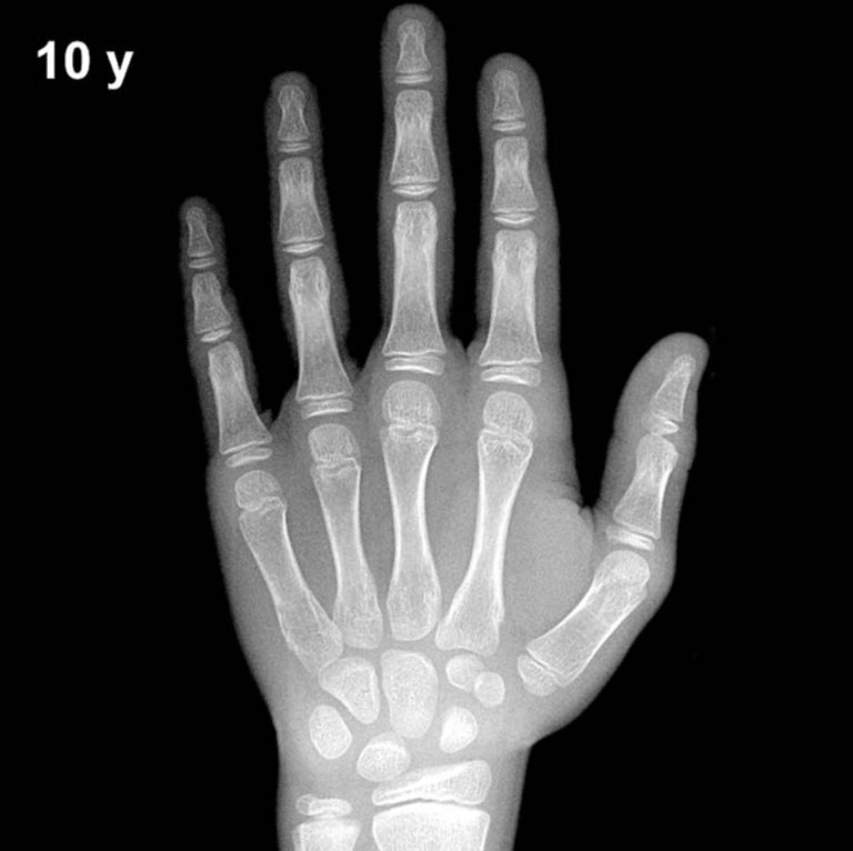
Bone Age in Girls Aged 18 Years — Greulich-Pyle Hand and Wrist X-Ray Reference
Bone age assessment using the Greulich-Pyle (GP) atlas compares a left-hand and wrist radiograph against standardized reference plates to estimate skeletal maturity. In 18-year-old girls, this assessment is particularly relevant for evaluating whether skeletal maturation is complete, with implications for growth potential, endocrine disorders, and medicolegal age estimation. The GP method remains the most widely used radiographic standard in pediatric and adolescent practice.
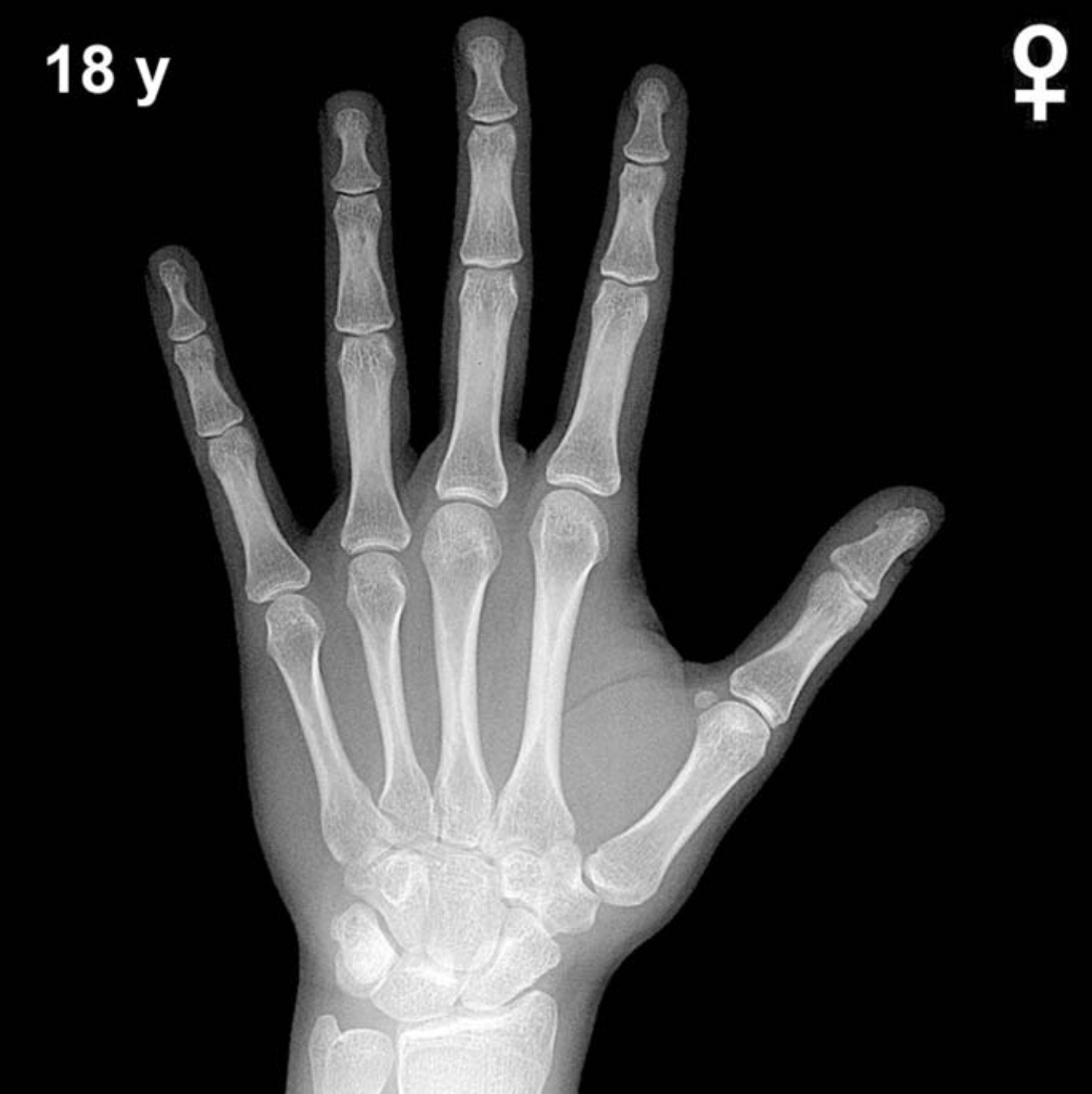
Expected Ossification Centers and Skeletal Findings
By 18 years of age in girls, skeletal maturation is typically complete or near-complete. The Greulich-Pyle atlas places the female standard for full skeletal maturity at approximately 17–18 years, meaning most girls at this age will demonstrate complete epiphyseal fusion across the hand and wrist.
- Distal radius and ulna: Epiphyses are typically fully fused to their respective metaphyses by this age; any visible growth plate represents delayed maturation.
- Metacarpal and phalangeal epiphyses: Complete fusion is expected at all rays; the middle and proximal phalangeal epiphyses fuse slightly earlier than distal ones in the female sequence.
- Carpal bones: All eight carpal ossification centers — capitate, hamate, triquetral, lunate, scaphoid, trapezium, trapezoid, and pisiform — are fully ossified and have reached adult morphology. The pisiform, which typically appears around 9–12 years in girls, is well-established by this stage.
- Sesamoid of the thumb (adductor pollicis): The ulnar sesamoid of the first MCP joint, a key peripubertal landmark appearing around Tanner stage 2–3, is fully ossified and mature.
The overall carpus and digit architecture at 18 years should reflect an adult skeletal pattern with no open physes visible on a properly exposed radiograph.
Clinical Pearls
At 18 years, a bone age concordant with chronological age confirms skeletal maturity and cessation of significant linear growth potential. A bone age substantially below 16 years in an 18-year-old girl warrants investigation for constitutional delay, hypothyroidism, growth hormone deficiency, or chronic systemic illness. Conversely, while advanced bone age is less clinically significant at this stage (maturation is already maximal), a history of markedly advanced bone age in earlier childhood may point to prior precocious puberty or exogenous androgen/estrogen exposure. Turner syndrome (45,X) is associated with delayed skeletal maturation and short stature, but is typically diagnosed well before age 18. A key interpretive pitfall is over-relying on a single radiograph without clinical context: ethnic and nutritional variation can shift bone age by ±1–2 years even in healthy individuals, and the GP atlas was standardized on a mid-20th-century North American cohort.
Reference: Greulich WW, Pyle SI. Radiographic Atlas of Skeletal Development of the Hand and Wrist. 2nd ed. Stanford University Press, 1959.