Bone Age in Girls Aged 14 Years — Greulich-Pyle Hand and Wrist X-Ray Reference
Bone age assessment using a left hand and wrist radiograph is a standard method for evaluating skeletal maturity in children and adolescents. The Greulich-Pyle atlas provides sex-specific standard radiographs against which a patient’s hand X-ray is compared to estimate skeletal age. At 14 years in girls, this assessment is particularly relevant in the workup of pubertal timing disorders, growth disturbances, and endocrine conditions.
Expected Ossification Centers and Skeletal Findings
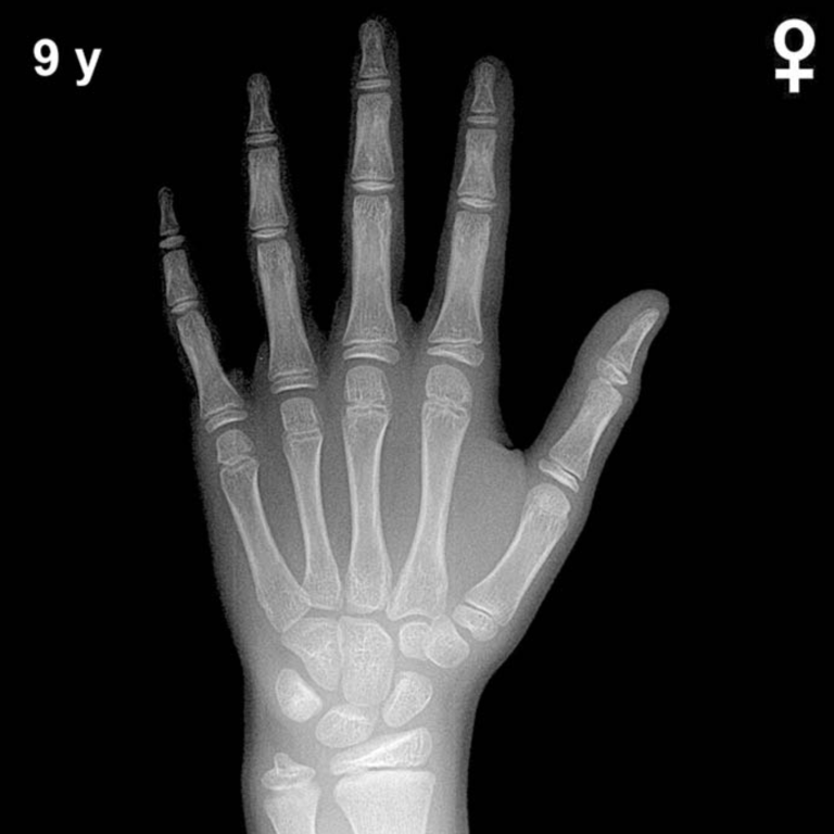
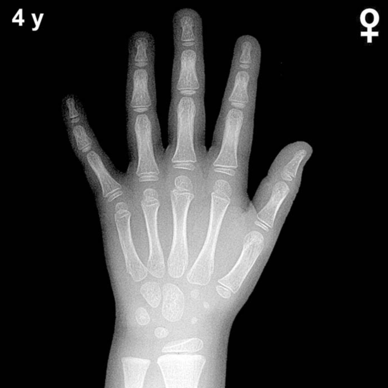
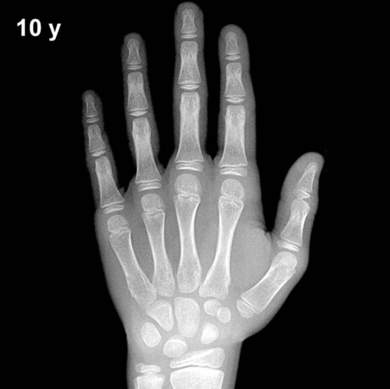
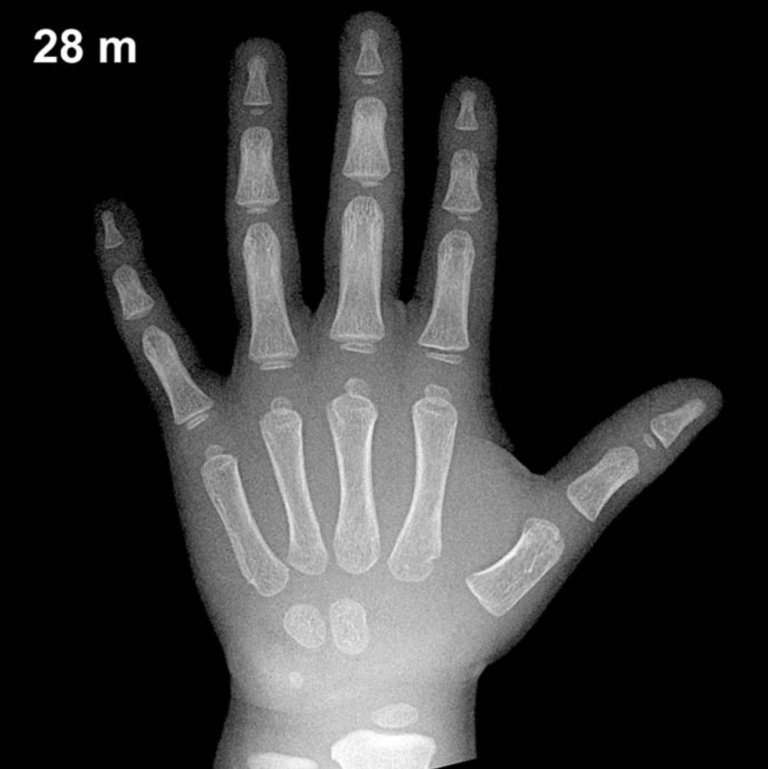
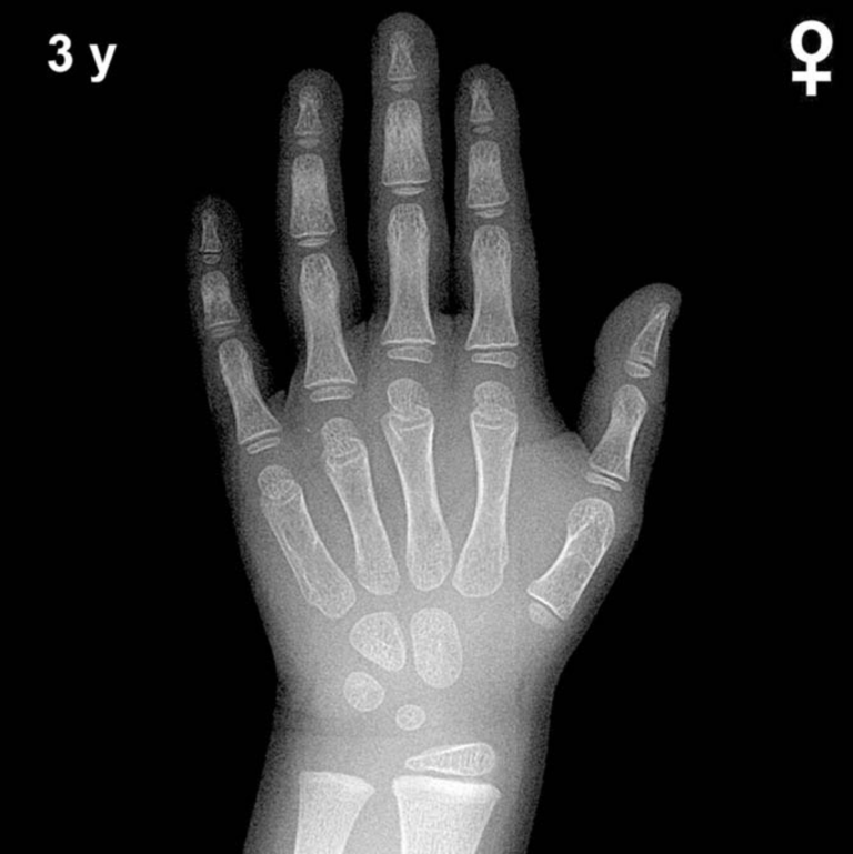
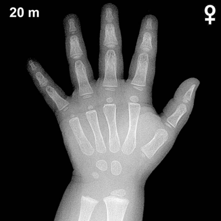
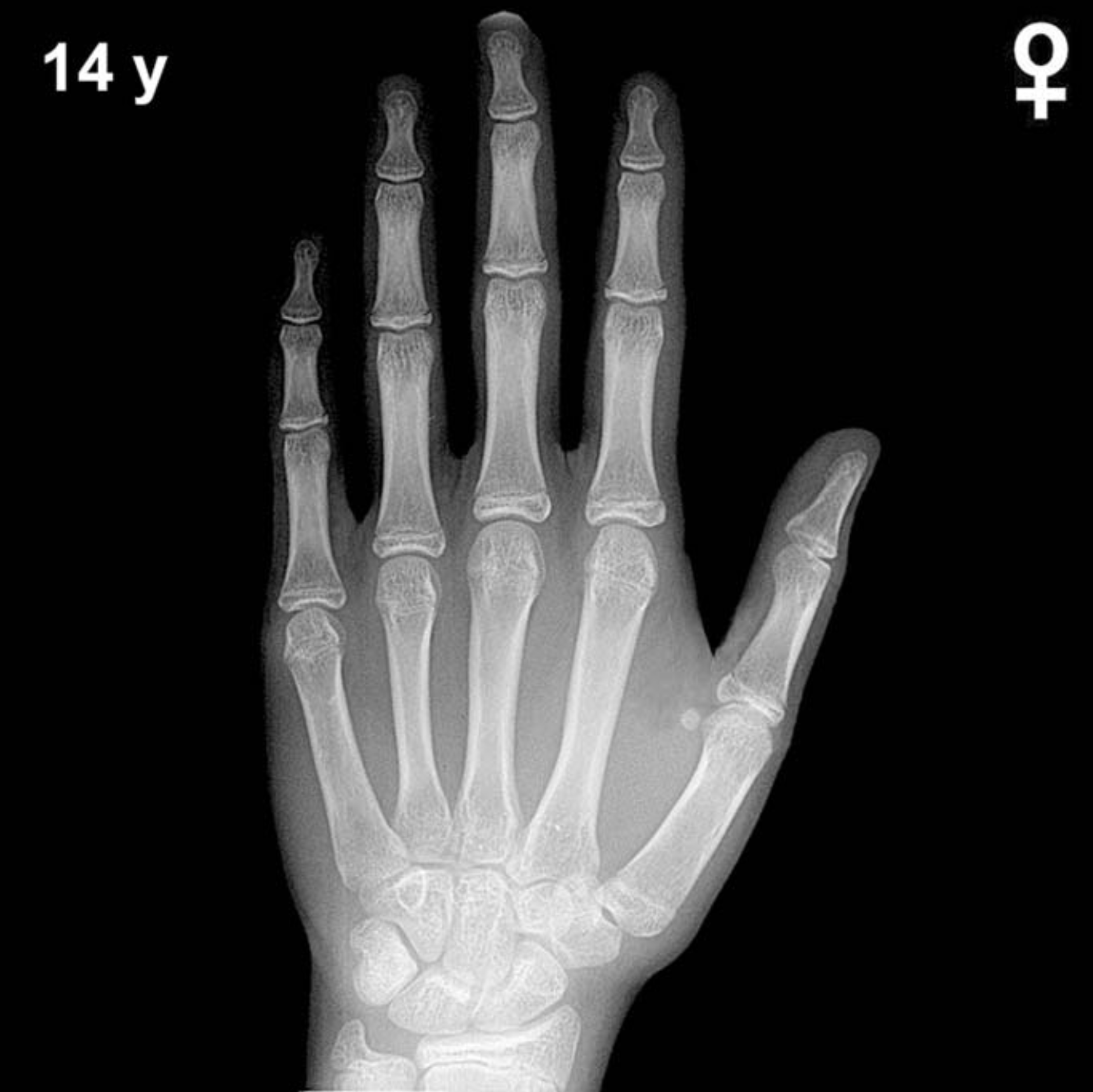
By 14 years of age in girls, all primary carpal bones are well ossified and have been present for several years. The capitate and hamate typically appear in early infancy, the triquetral by 2–3 years, the lunate by 3–4 years, the scaphoid, trapezium, and trapezoid by 4–6 years, and the pisiform typically by 9–12 years in girls — and should be clearly visible by this age. All carpal ossification centers are therefore expected to be present and well developed.
At 14 years, the epiphyses of the distal radius and distal ulna are well formed and substantially enlarged, with the distal radial epiphysis typically showing progressive capping of the metaphysis. In girls, epiphyseal fusion at the phalanges and metacarpals is an important landmark; by approximately 13–15 years, fusion of the proximal and middle phalangeal epiphyses may be commencing or partially complete in some individuals. The adductor sesamoid of the thumb, a key peripubertal marker, is typically present by 13–14 years in girls and should be visible at this age. The distal radial and ulnar physes remain open but are narrowing as skeletal maturity approaches.
- All carpal bones ossified and well developed
- Pisiform present and well formed
- Adductor sesamoid of the thumb expected to be present
- Distal radial and ulnar epiphyses enlarged; physes narrowing
- Early or partial epiphyseal fusion at phalanges possible
Clinical Pearls
Girls are skeletally advanced compared to boys by approximately 1–2 years throughout childhood and adolescence, a difference well documented in the Greulich-Pyle atlas. At 14 chronological years, a bone age significantly advanced beyond 15 years may raise concern for precocious puberty or exogenous androgen/estrogen exposure, while a bone age delayed below 12 years warrants evaluation for growth hormone deficiency, hypothyroidism, constitutional delay of growth and puberty, or Turner syndrome. Standard deviation for bone age at this age is approximately ±1 year, so mild discrepancies are within the expected range of normal variation.
A key interpretive pitfall is over-reliance on a single skeletal indicator; bone age should always be assessed as a composite across multiple bones rather than any single epiphysis. Population differences in skeletal maturation may also limit direct applicability of the Greulich-Pyle standards, which were derived from a mid-20th century North American cohort. Reference: Greulich WW, Pyle SI. Radiographic Atlas of Skeletal Development of the Hand and Wrist. 2nd ed. Stanford University Press, 1959.