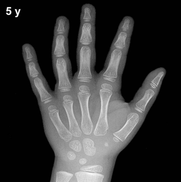
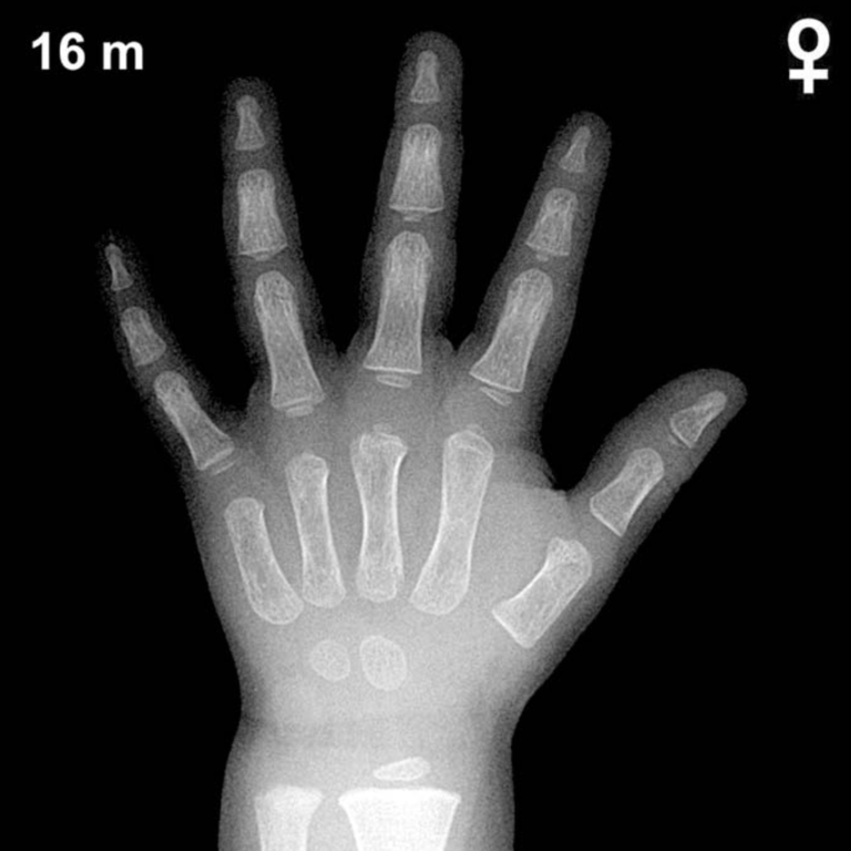
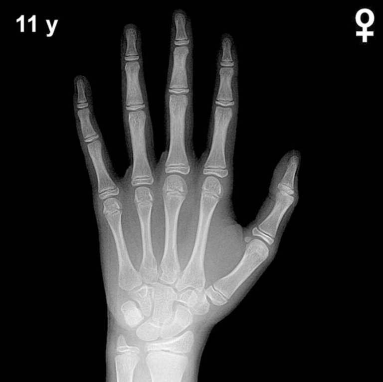
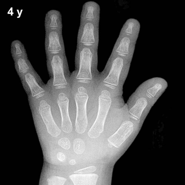
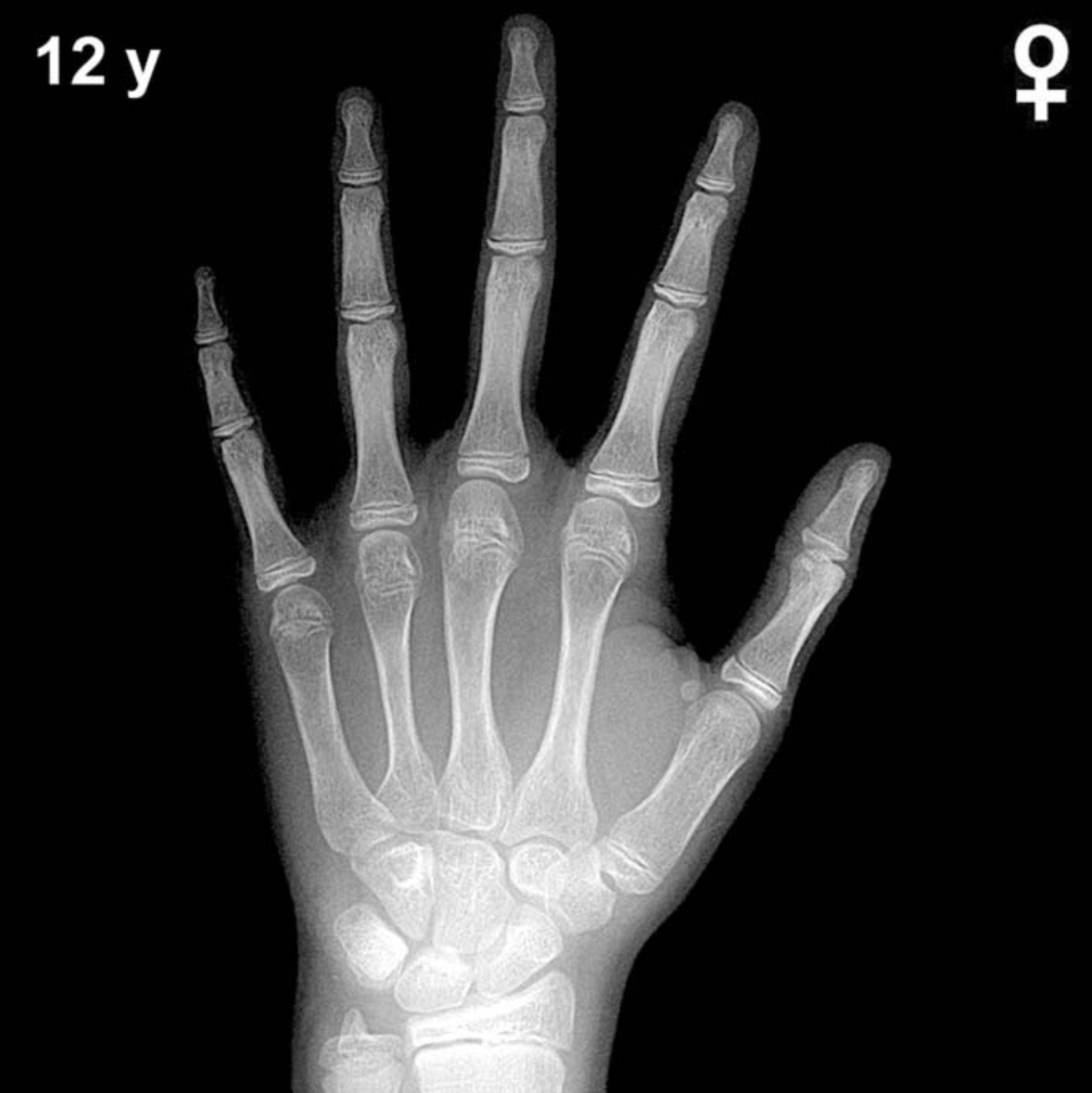
Bone Age in Girls Aged 12 Years — Greulich-Pyle Hand and Wrist X-Ray Reference
Bone age assessment using a posteroanterior hand and wrist radiograph is a well-established method for evaluating skeletal maturity in children and adolescents. The Greulich-Pyle (GP) atlas provides sex-specific standard radiographs against which a patient’s film is compared to derive a skeletal age. In 12-year-old girls, this assessment is particularly relevant for investigating precocious or delayed puberty, growth hormone disorders, and monitoring conditions that affect skeletal maturation.
Expected Ossification Centers and Skeletal Findings
By 12 years of age in girls, all eight carpal bones are typically well ossified. The capitate and hamate, the earliest to appear (at approximately 3 and 6 months of life, respectively), are now mature in size and morphology. The triquetral, lunate, scaphoid, trapezium, and trapezoid are all expected to be present and well developed. The pisiform, which appears later — typically around 9–12 years in girls — should be visible or just emerging at this age, making its presence or absence a useful maturational marker at this stage.
Epiphyseal development is advanced in 12-year-old girls, reflecting proximity to or active progression through puberty. The distal radial epiphysis is well established and typically shows significant capping of the metaphysis. The distal ulnar epiphysis, which appears around 5–7 years, is now well formed. Epiphyses of the metacarpals and phalanges are broad and may show early or progressive fusion in more skeletally advanced individuals. The adductor sesamoid of the thumb, a key peripubertal landmark, typically appears around 11–13 years in girls and is expected to be present or appearing at this chronological age.
- All 8 carpal bones: ossified and well developed
- Pisiform: present or just appearing (~9–12 yr in girls)
- Distal radial and ulnar epiphyses: well formed, with metaphyseal capping
- Thumb adductor sesamoid: typically present at this age in girls
- Phalangeal and metacarpal epiphyses: broad; early fusion may be seen in advanced individuals
Clinical Pearls
Girls are skeletally more mature than boys of the same chronological age by approximately 1–2 years across childhood and adolescence, a difference well documented in the Greulich-Pyle atlas. At 12 years, a normal range of skeletal maturity spans roughly 10–14 years, reflecting the wide physiological variability of pubertal timing. A bone age significantly advanced beyond 13–14 years in a 12-year-old girl should prompt evaluation for precocious puberty or exogenous androgen/estrogen exposure, while a bone age below 10 years warrants consideration of growth hormone deficiency, hypothyroidism, or constitutional delay of growth and puberty. In girls with Turner syndrome (45,X), bone age is often only mildly delayed but growth velocity is impaired, and the clinical context is essential.
A key interpretive pitfall is inter-observer variability: GP atlas comparisons can vary by up to 6–12 months between readers. Standardized training and, where available, digital bone age tools can improve reproducibility. Clinical correlation with growth charts, pubertal staging, and hormonal data remains essential. Reference: Greulich WW, Pyle SI. Radiographic Atlas of Skeletal Development of the Hand and Wrist. 2nd ed. Stanford University Press, 1959.