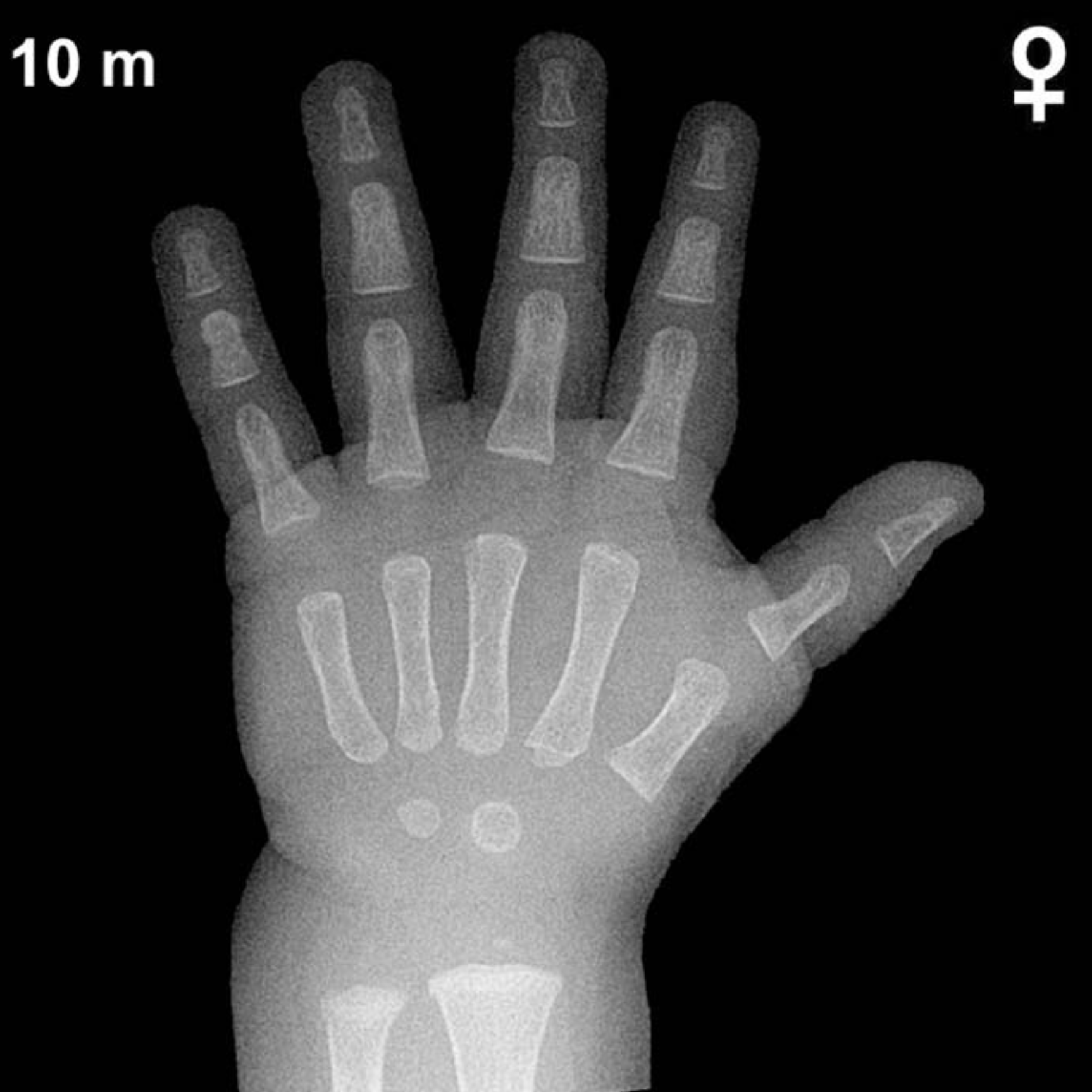
Bone Age in Girls Aged 10 Months — Greulich-Pyle Hand and Wrist X-Ray Reference
Bone age assessment using the Greulich-Pyle (GP) atlas involves comparing a left hand and wrist radiograph to standardized reference plates to estimate skeletal maturity. In infants and toddlers, this evaluation is particularly relevant when investigating growth faltering, suspected endocrine disorders, or when estimating developmental age in uncertain birth history situations. Early identification of skeletal maturation discrepancies can prompt timely workup for conditions such as hypothyroidism or growth hormone deficiency.
Expected Ossification Centers and Skeletal Findings
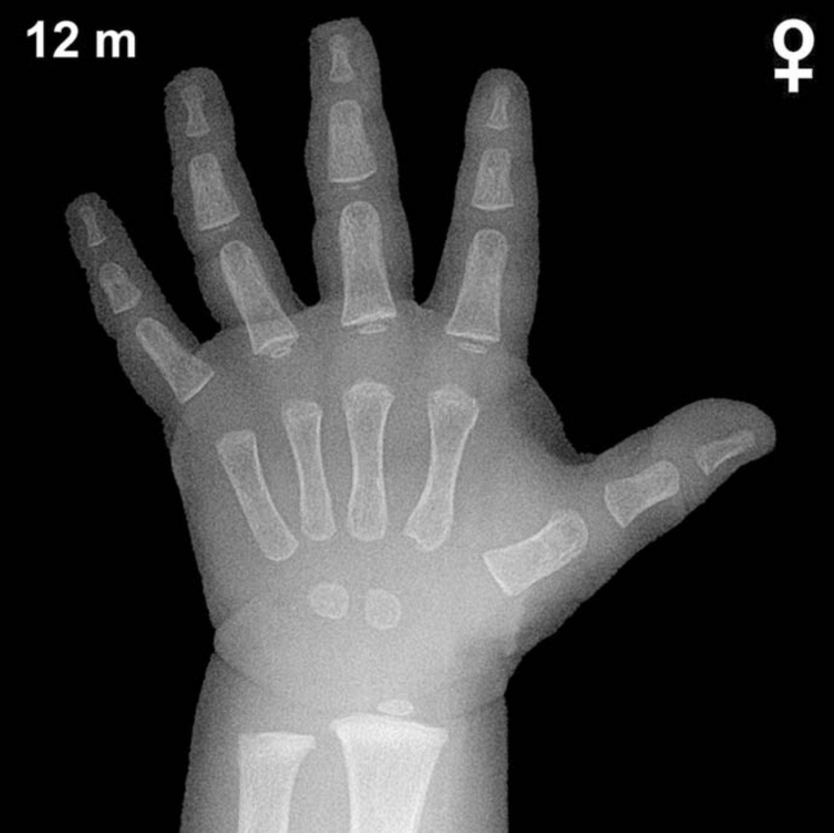
At 10 months of age in girls, the Greulich-Pyle atlas anticipates a limited but important set of ossification centers in the hand and wrist. The capitate and hamate are the earliest carpal bones to ossify, typically appearing by approximately 2–6 months of age; both should be well-visualized and of reasonable size by 10 months. The distal radial epiphysis typically becomes visible around 9–12 months in girls, so its presence or absence at this age is a critical landmark to assess.
- Capitate: present and well-formed by this age
- Hamate: present by this age, often with a visible body
- Distal radial epiphysis: typically just appearing or early in development at 10 months in girls
- Triquetral, lunate, scaphoid, trapezium, trapezoid, pisiform: not yet expected at this age
- Distal ulnar epiphysis: not yet expected; typically appears around 5–7 years
- Metacarpal and phalangeal epiphyses: may show early, subtle ossification in the more mature infant
Overall carpal bone count at this age should be two (capitate and hamate). The appearance of additional carpal centers or a well-developed distal radial epiphysis would suggest skeletal advancement beyond chronological age.
Clinical Pearls
Skeletal maturation in girls is generally ahead of boys by approximately 1–2 months at this very young age, a gap that widens significantly during later childhood and puberty. At 10 months, normal bone age variability spans roughly ±2–3 months around the chronological age. A bone age markedly delayed relative to chronological age may raise concern for hypothyroidism, hypopituitarism, or severe malnutrition, while accelerated skeletal maturity warrants consideration of precocious puberty or excess androgen exposure. A key interpretive pitfall at this age is over-relying on a single ossification center: the distal radial epiphysis can be subtle and positioning-dependent, so clinical correlation and serial imaging are recommended when findings are borderline. Reference: Greulich WW, Pyle SI. Radiographic Atlas of Skeletal Development of the Hand and Wrist. 2nd ed. Stanford University Press, 1959.